The 25 most jaw-dropping images from Nikon’s Small World 2023 microphotography contest
Take a very close look at alien spiders, teeny cuckoo wasps, pork parasites and caffeine crystals.
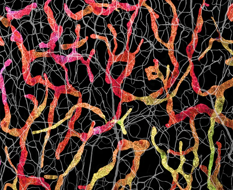
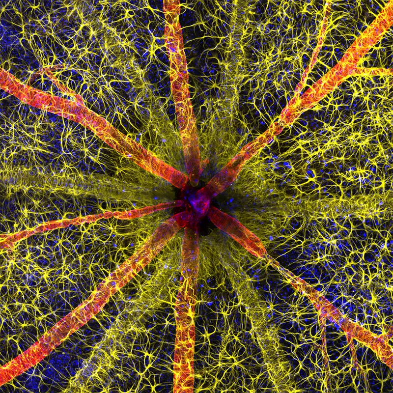
Rodent optic nerve head showing astrocytes (yellow), contractile proteins (red) and retinal vasculature (green). Photo by Hassanain Qambari & Jayden Dickson/Nikon Small World
The winners of the most prestigious prize in science imaging have just been announced, with an incredible image of an optic nerve of a rat taking the title.
The image, which was taken by Hassanain Qambari and assisted by Jayden Dickson of the Lions Eye Institute, provides an important contribution to the study of diabetic retinopathy, an eye disease which affects one in five persons with diabetes worldwide.
Diabetic retinopathy occurs when high blood sugar damages the blood vessels in the tissue at the back of the eye, known as the retina. The damaged blood vessels can swell and leak, which causes blurry vision and total loss of eyesight in some cases. Since 2021, Qambari has devoted his time and research to the early detection and reversal of the disease.
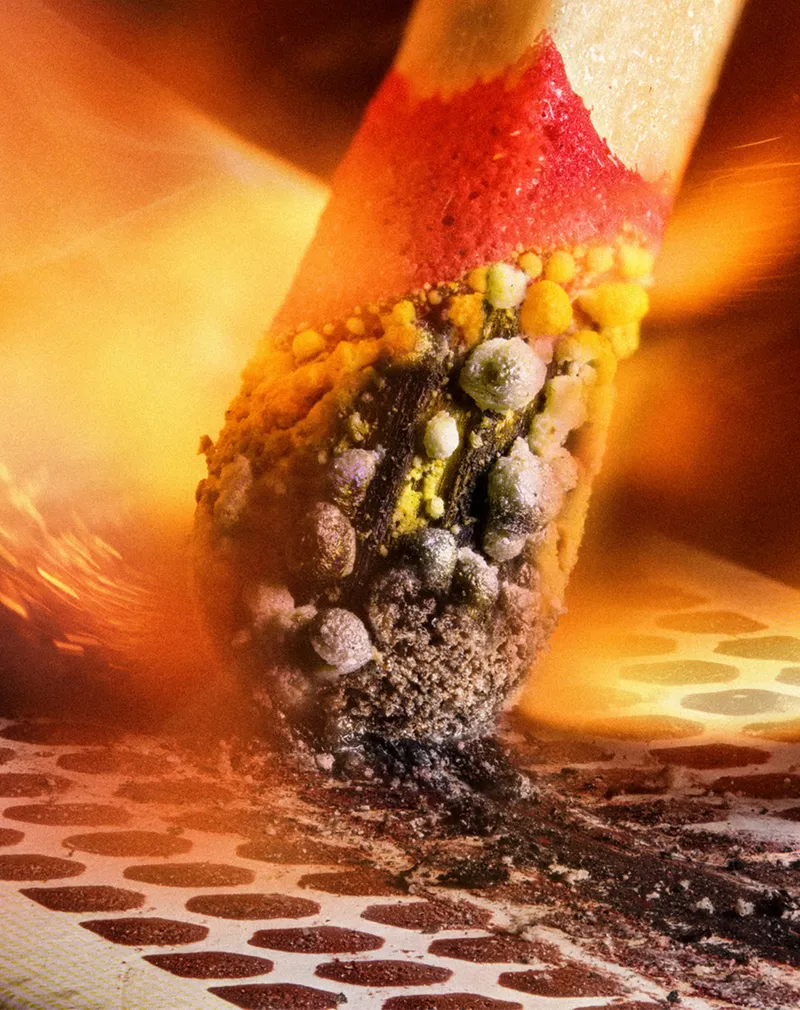
Second place was awarded to Ole Bielfeldt for his dramatic image of a matchstick igniting by the friction surface of the box. The image was taken within one eight-thousands of a second and is made up of multiple images.
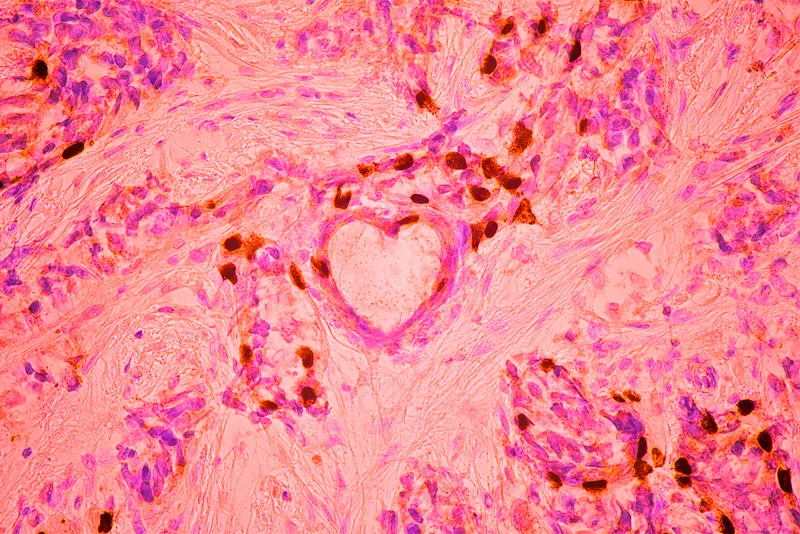
Third placewas awarded to Malgorzata Lisowska for her image of breast cancer cells.
Now in its 49th year, Nikon Small World is open to anyone with an interest in photography or video. It is renowned for celebrating photographic and artistic excellence in photomicrography.
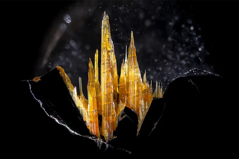
Image of Distinction
Rutilated quartz like the one pictured is a variety of quartz which contains needle-like inclusions. Photo by Danny Sanchez/Nikon Small World
Image of Distinction
A crab spider (Thomisus onustus) photographed at 6.3X magnification. Photo by Sébastien Malo/Nikon Small World
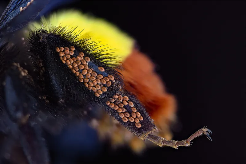
Image of Distinction
Phoretic mites on the leg of a bumblebee, photographed at 3X magnification. Phoresy is an interaction in which a creature such as a mite attaches itself to a host animal for the purpose of travel. Photo by Amir Maqbool/Nikon Small World
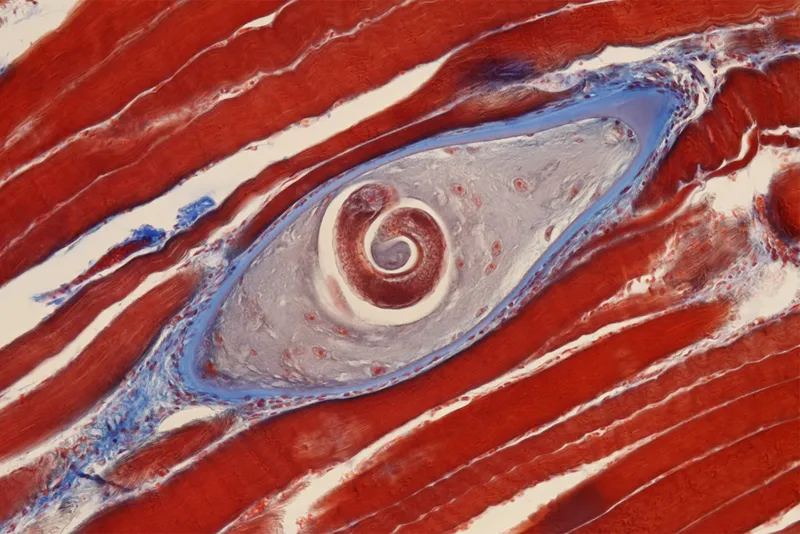
Honourable mention
Trichinella, as seen within pork tissue. Trichinella is a parasitic worm known to cause trichinosis, which can cause severe symptoms in the human digestive system if the meat is not cooked properly. Photo by Dr Nathan P Myhrvold/Nikon Small World
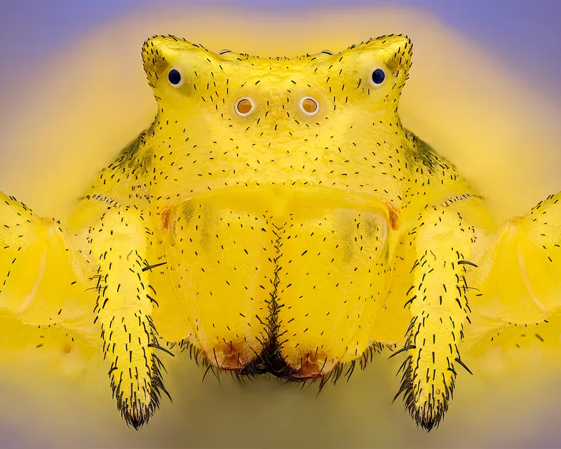
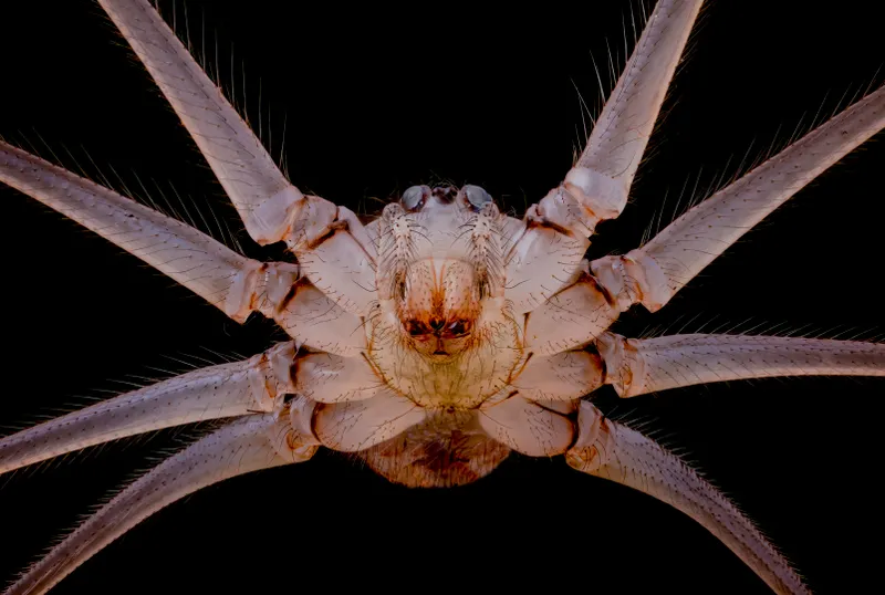
Honourable mention
The underside of cellar spider (Pholcus phalangioides) at 10X magnification. Photo by Dr Andrew M Posselt/Nikon Small World
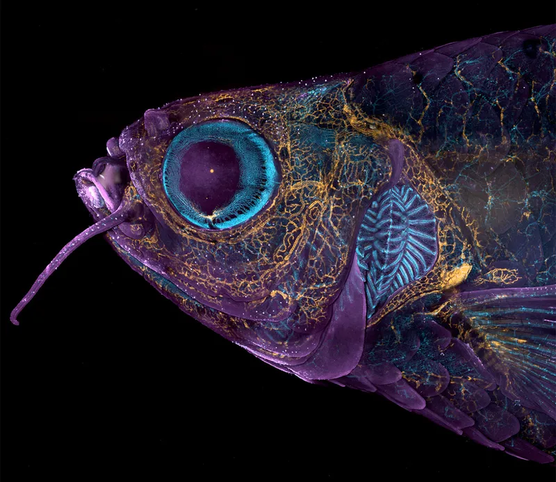
Top 20
An adult transgenic zebrafish head showing blood vessels in blue, lymphatic vessels in yellow, and the skin and scales in magenta. Photo by Daniel Castranova & Dr Brant Weinstein/Nikon Small World
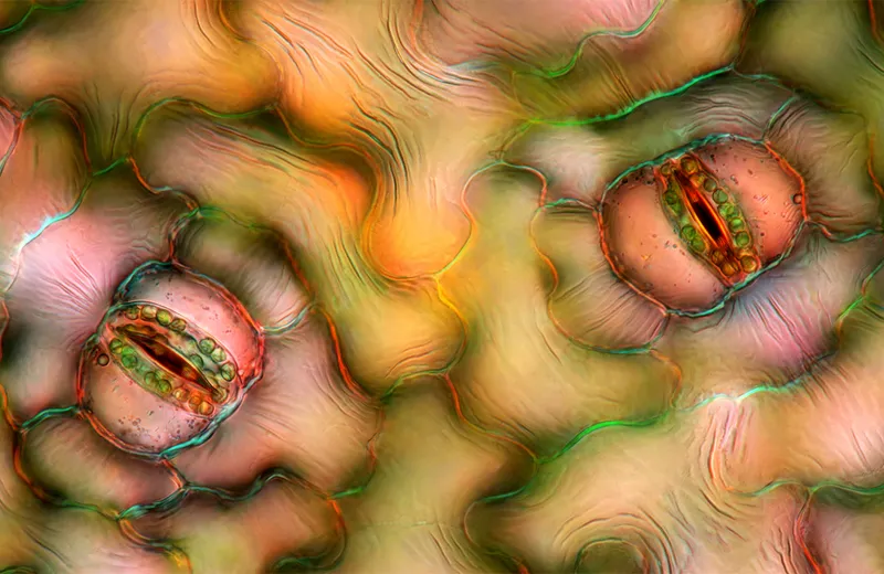
Top 20
A stomata in a peace lily (Spathiphyllum sp.) leaf epidermis. Photo by Marek Miś/Nikon Small World
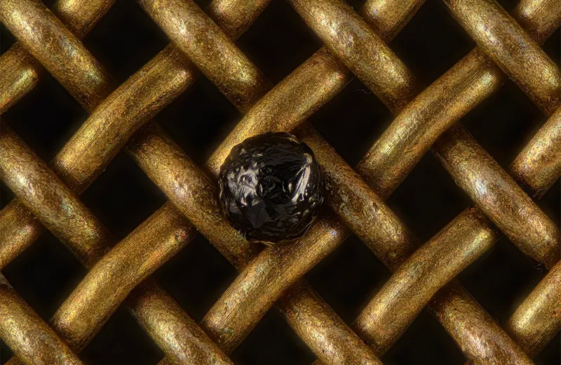
Top 20
A cryptocrystalline micrometeorite resting on a testing sieve. Photo by Scott Peterson/Nikon Small World
Top 20
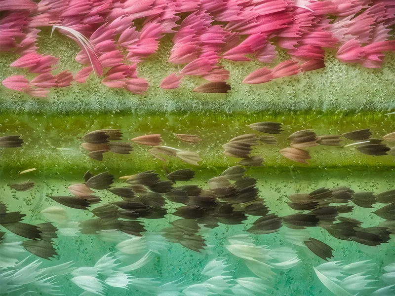
Chinese moon moth (Actias ningpoana) wing scales at 20X magnification. Photo by Yuan Ji/Nikon Small World
Top 20
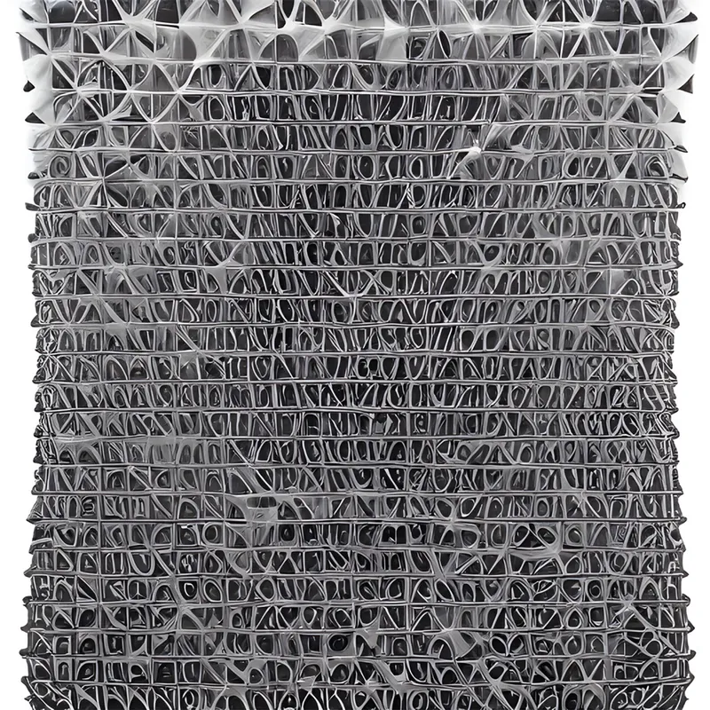
Carbon nanotubes photographed at 30X magnification. Photo by Dr Diego García/Nikon Small World
Top 20
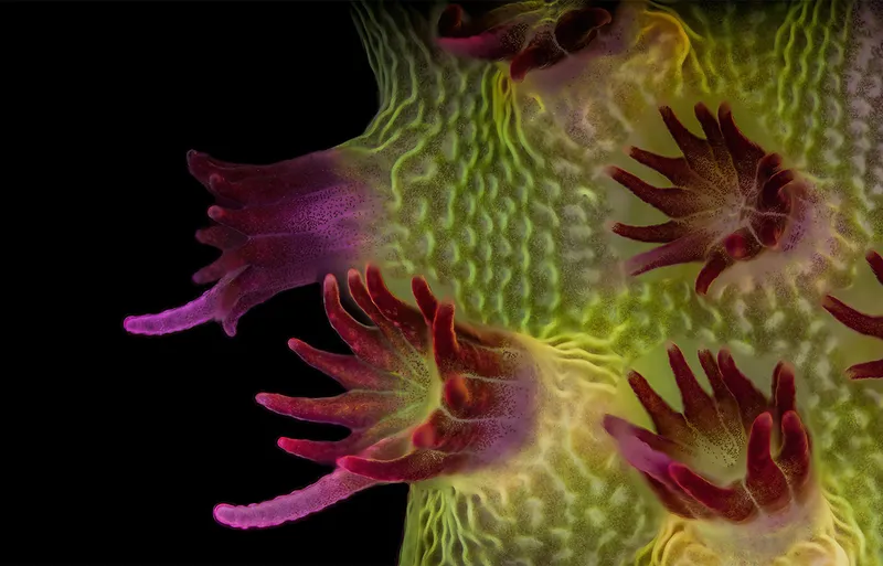
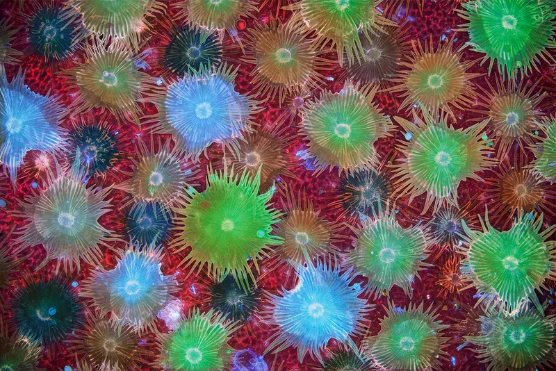
Fluorescent image of a stony coral (Acropora sp.) showing individual polyps with symbiotic zooxanthellae. Photo by Dr Pichaya Lertvilai/Nikon Small World
Top 20
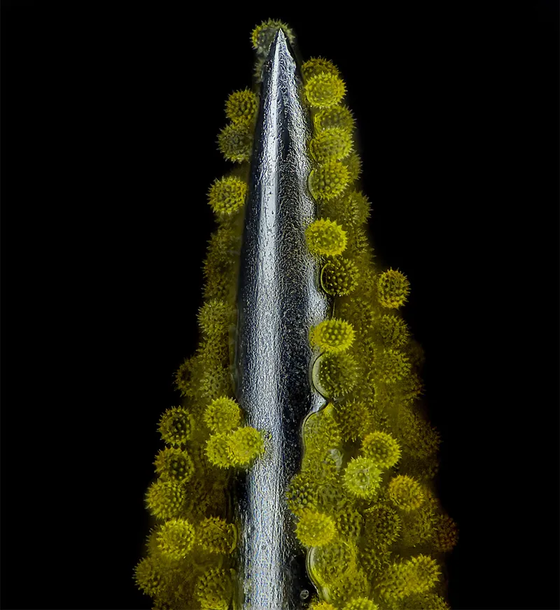
Sunflower pollen on an acupuncture needle, photographed at 40X magnification. Photo by John-Oliver Dum/Nikon Small World
Top 20
Blood and lymphatic vasculatures in the ear skin of an adult mouse. Photo by Satu Paavonsalo & Dr Sinem Karaman/Nikon Small World
Top 20
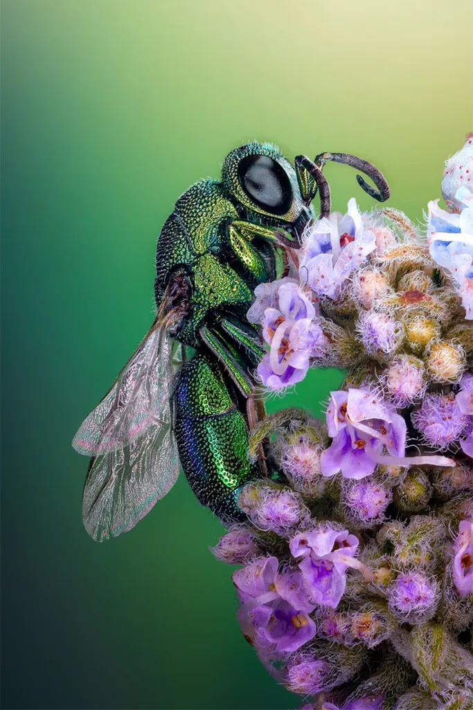
A cuckoo wasp (Chrysididae) standing on a flower in Tanta, Egypt. This species in known for bright colours, and lay their eggs in the nests of unrelated species. Photo by Sherif Abdallah Ahmed/Nikon Small World
Top 20
Crystallised sugar syrup imaged at 25X magnification. Photo by Dr Diego García/Nikon Small World
Top 10
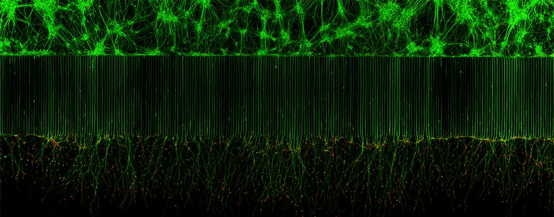
Motor neurons grown in a microfluidic device for the separation of cell bodies (top) and axons (bottom). The green lines are microtubules. Photo by Melinda Beccari & Dr Don W Cleveland/Nikon Small World
Top 10
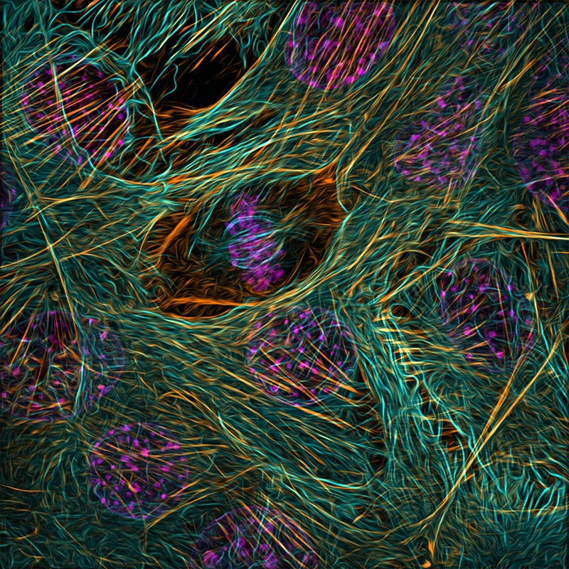
Cytoskeleton of a dividing myoblast showing tubulin (cyan), F-actin (orange) and nucleus (magenta). Photo by Vaibhav Deshmukh/Nikon Small World
Top 10
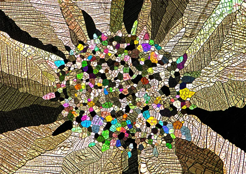
Polarised light image of caffeine crystals at 25X magnification. Photo by Stefan Eberhard/Nikon Small World
Top 10
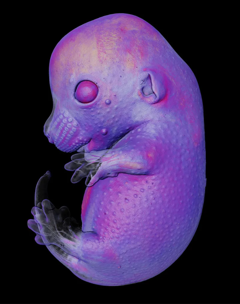
A mouse embryo imaged at 4X magnification. Photo by Dr Grigorii Timin & Dr Michel Milinkovitch/Nikon Small World
Top 10
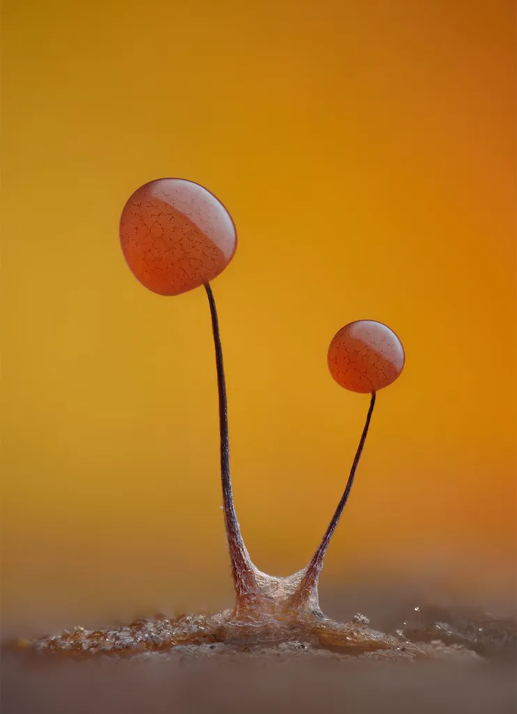
A slime mould (Comatricha nigra) showing capillitial fibers through its translucent peridium. Taken at 10X magnification. Photo by Timothy Boomer/Nikon Small World
Auto-fluorescing defensive hairs covering the leaf surface of a wild olive tree (Eleagnus angustifolia) exposed to UV light. Photo by Dr. David Maitland/Nikon Small World
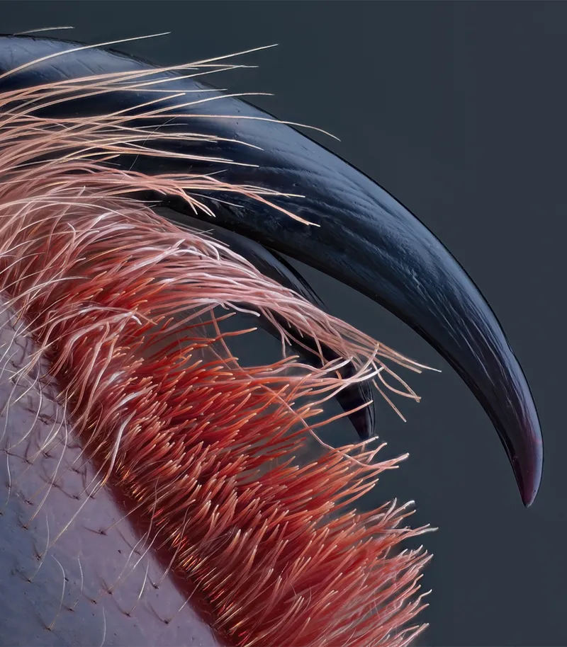
Top 10
Venomous fangs of a small tarantula at 10X magnification. Photo by John-Oliver Dum/Nikon Small World
Third place
Brightfield image of breast cancer cells at 40X magnification. Photo by Malgorzata Lisowska/Nikon Small World
Second place
Brightfield image of a matchstick igniting by the friction surface of the box at 2.5X magnification. Photo by Ole Bielfeldt/Nikon Small World
Overall winner
Rodent optic nerve head showing astrocytes (yellow), contractile proteins (red) and retinal vasculature (green). Photo by Hassanain Qambari & Jayden Dickson/Nikon Small World
James Cutmore is the picture editor of BBC Science Focus Magazine. He has worked on the magazine and website for over a decade, telling compelling science stories through the use of striking imagery. He holds a degree in Fine Art, and has been nominated for the British Society of Magazine Editors Talent Awards, being highly commended in 2020. His main areas of interest include photography that highlights positive technology and the natural world. For many years he was a judge for the Wellcome Trust's image competition, as well as judging for the Royal Photographic Society.