The British Heart Foundation (BHF) has just announced the winner of its annual 'Reflections of Research' photography competition. The competition challenges BHF-funded scientists to showcase their heart research through the use of amazing images.
The winning image 'Paths of the heart' was taken by Dr Marina Strocchi, and shows a map of muscle cells in the human heart.
“In our research, computer models help doctors understand so much more about diseases of the heart. By simulating how electrical activity spreads through the heart, following the direction shown in the picture, to then trigger a heartbeat, we can predict how specific patients will respond to treatments to personalise and adjust how we care for them" said Dr Strocchi.
The BHF funds ground-breaking research that aims to eradicate heart and circulatory diseases around the world.
Shortlisted – A broken heart

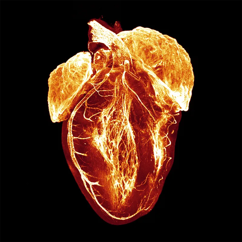
What could be two intricate butterfly wings are in fact the two halves of a rabbit heart in recovery, several weeks after a heart attack. A powerful MRI scanner was used to build a 3D representation of the heart and split it in two to reveal the inner structure in amazing detail. The upper and lower chambers of the heart, the heart muscle, and the network of fibres inside the chambers can all be seen.
People who have had a heart attack are more at risk from dangerous heart rhythms and sudden death. This may be because of scarring of the heart tissue after an attack. By studying how scarring interacts with the heart muscle and the electrical signals in the heart, Michael Freeman (University of Glasgow) hopes to find out why this risk increases. This could allow doctors to identify those at high risk of a heart rhythm problem.
Shortlisted – Have a heart
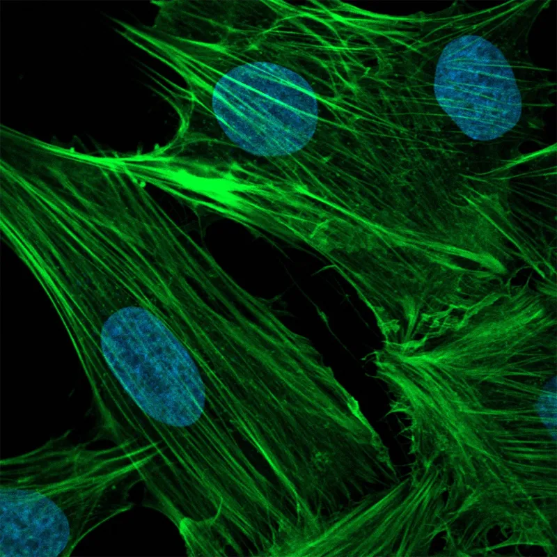
In this image, the web of fibres and deep blue circles show different parts of heart muscle cells dyed with fluorescent molecules. Each circle is the nucleus of a cell, the control centre that contains the genetic information. Each green tendril is a filament of the protein actin, part of the machinery that allows the heart to contract during the heartbeat to push blood out and around the body.
Shortlisted – A close-up view of vascular first aid kits
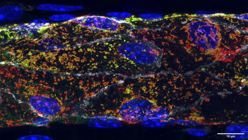
The image shows a blood vessel in minute detail. The white streaks are the edges of the cells that make up the lining of the blood vessel, known as endothelial cells. Each blue oval is the genetic information at each cell’s centre. The constellation of green, yellow, and red points are storage sites for von Willebrand factor (VWF), a molecule that the cells release to create blood clots when the vessel is damaged.
Shortlisted – Development and Remodelling of The Human Fetal Valves
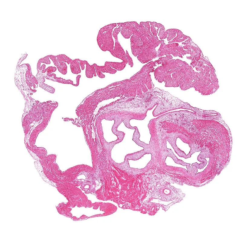
The colours and patterns in this image show the human heart after 12 weeks of development. Congenital heart disease affects at least one in every 150 live births in the UK. The knowledge gained by observing hearts develop is being combined with the vast amount we already know about CHD development in mice. The researchers think that a better understanding of how CHDs develop can lead to targeted treatments for the conditions.
Shortlisted – A window into the heart of artificial intelligence
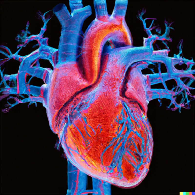
This bright neon representation of a heart has actually been created by the AI image generator Dall-E, from the sentence 'An image of the heart from a computed tomography scan, digital art'. Whilst able to create a stunning image, the AI has made a few mistakes in the anatomy and structure of this heart, most noticeably in the branching pattern of the blood vessels.
Shortlisted – The glue that holds your life together
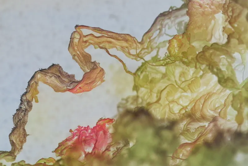
This picture shows the inside of the aorta, the largest blood vessel in the human body, taken at 8000X magnification using an electron microscope. In it, we can see the individual fibres of collagen within the walls of the blood vessel. This protein acts like glue in the wall of the blood vessels, giving them the strength needed to withstand the high pressure of blood as it is pumped around the body.
A swelling or building of the blood vessel is called an aneurysm. If the blood vessel gets too large it can burst, leading to internal bleeding. Ruptures of the aorta are responsible for over 4,000 deaths a year in the UK. The team at the University of Leicester hopes to find ways of preventing this by studying how collagen differs in different areas of the aorta wall.
Shortlisted – The radiant microvascular realm
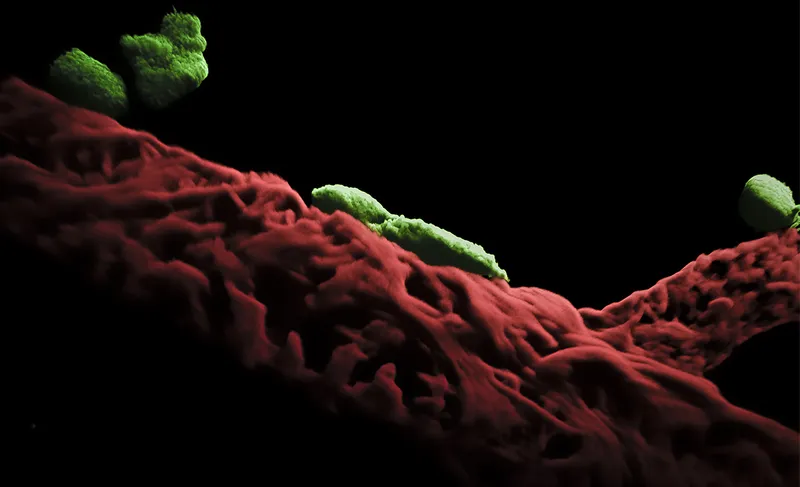
The red mass at the bottom of the picture are cells on the outside of a mouse’s blood vessel. The green bodies attached like leeches are called mast cells, a type of immune cell responsible for controlling blood flow and inflammation.
Research by Dr Loïc Rolas focuses on the interaction between blood vessels and immune cells, the closeness of which is shown by this image. He has discovered a number of ways in which this close relationship becomes more dysfunctional as we age.
Shortlisted – The embryonic human heart
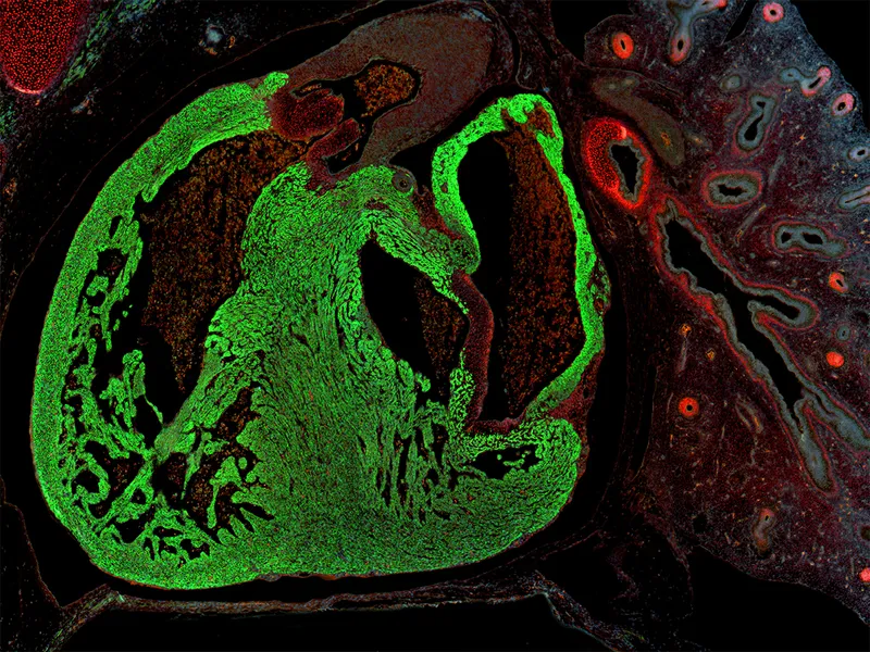
This is a side view of a human heart and lung shown after 56 days of development. At this stage, the heart is only 2mm in size. Using a fluorescent microscope we can see the boundaries of the heart muscle (green) and the heart valves (red).
Supporter's Favourite – Seeing through the heart
Fantastic bursts of light in this image highlight blood vessels around the outside of the heart and areas of the electrical system at its centre. This system is visible as the tangle of fibres in the middle of the image.
The two larger glowing areas are the atria – the top two chambers of the heart – and the branch around the outside on the left of the image, is a coronary artery, which supplies blood to the heart muscle.
Overall Winner – Paths of the heart
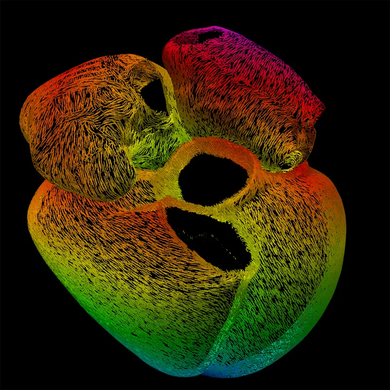
This multicoloured structure, generated by a computer model, is a map of bundles of cells that together make up the heart muscle. These bundles, each shown by a separate line, transmit the electrical signal that causes the heart to beat.
Each line in the image indicates the direction the electrical signal travels. The signal starts in the two top chambers of the heart (the atria) and moves to the bottom two (the ventricles).
Read more: