Moments of pure eureka: 10 best photos of recent medical breakthroughs
Check out the latest winners from Great Ormond Street Hospital's latest photography competition, detailing stunning breakthroughs in science.
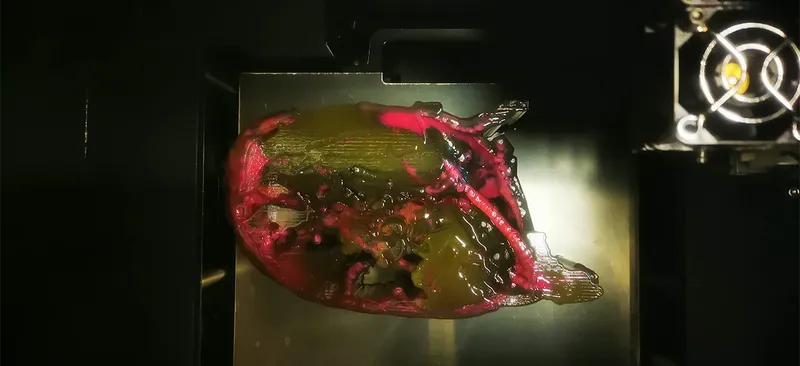
This image shows a gut ‘mini-organ’, known as an organoid. During a process used to visualise specific proteins, one of the organoids exploded, revealing its inner workings. Tiny organs like this one can be used to model gastrointestinal diseases, and are the perfect tool for scientists to study new therapies and test new drugs in the laboratory. Photo by Giada Benedetti/GOSH
The history of science is full of those ‘eureka!’ moments when a theory is proved or a breakthrough is made. And it is those kinds of breakthroughs that are being celebrated in the Great Ormond Street Hospital’s latest image competition, ‘A Moment of Discovery’.
Staff from across Great Ormond Street Hospital for Children NHS Foundation Trust (GOSH) and its affiliated institutes submitted images that captured an important milestone in their research area. The three most popular shortlisted images amongst a panel of expert judges were selected and put to a public vote via social media.
The images that made the shortlist ranged from vibrant microscopy to cartoon illustrations. These images offer glimpses into the research being undertaken at GOSH. Through this research, it is hoped that new treatments for rare and complex conditions can be found to help transform the lives of seriously ill children and young people.
The winning image was taken by Giada Benedetti, a PhD student working at the Zayed Centre for Research into Rare Disease in Children (ZCR). This striking image shows a gut organoid (or 'mini-organ') as it explodes, revealing its inner workings.
Organoids are minuscule 3-dimensional tissue cultures that are derived from stem cells and can be tailored to replicate many different types of organs in the human body, such as the heart or liver.
In celebration of Rare Disease Day on the 29th of February, all of the winning and shortlisted images will be exhibited at an event in the Zayed Centre for Research into Rare Disease in Children.
Shortlisted – My lung is on fire
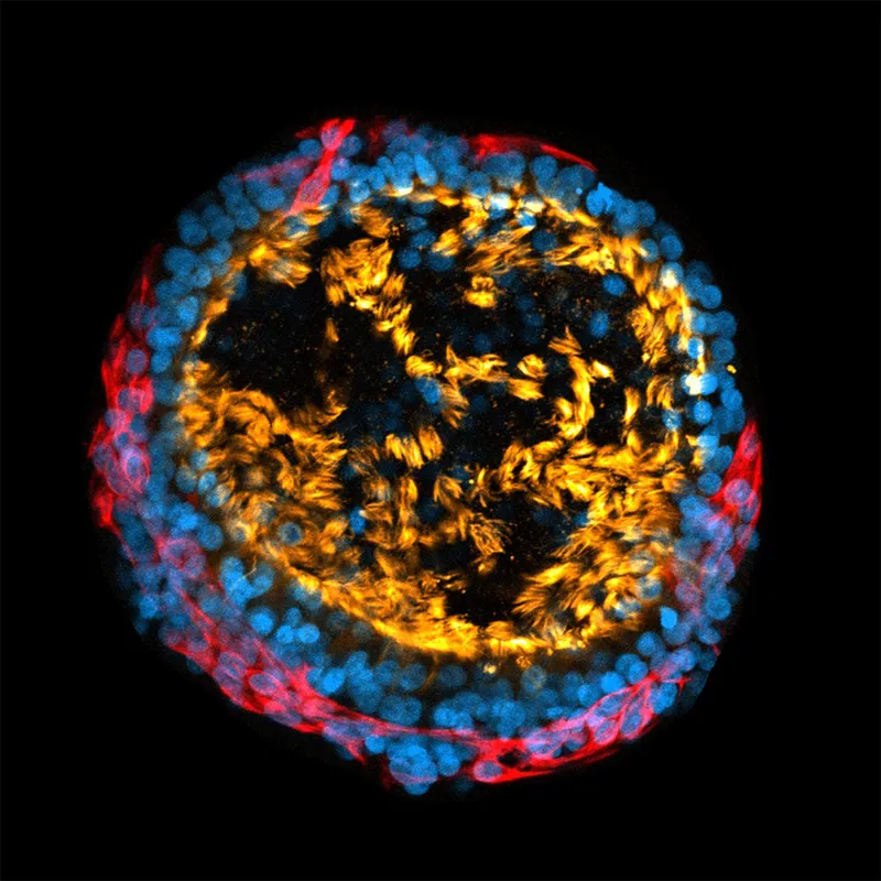
This lung ‘mini-organ’, known as an organoid, was grown from stem cells. It is a tiny, functional replica of a lung, mirroring its complexity. The blue shows the cell nuclei, the red shows the cell membrane, and the yellow/orange 'fire' shows the moving hair-like structures called cilia. Photo by Giuseppe Cala/GOSH
undefined
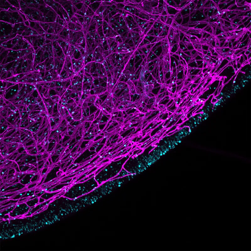
Shortlisted – Glial cells in action
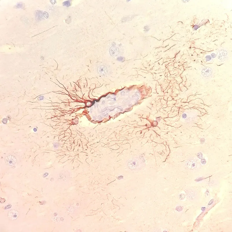
This image shows nerve helper cells known as glial cells (shown in brown), which have long fibre-like structures that adhere to blood vessels and transport nutrients and oxygen to surrounding nerve cells. Photo by Lucien Bonfante/GOSH
Shortlisted – The lights of life
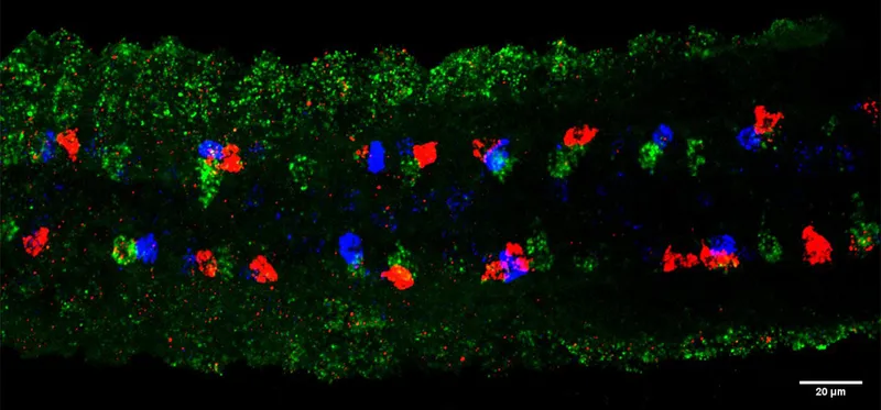
The spinal cord of a zebrafish embryo with different neurons produced via asymmetric division can be seen in this image. Confocal imaging identified the progenitor cells (green) that produce two distinct daughter cells (red and blue) via asymmetric division. Photo by Atachapon Theppichaiyanond/GOSH
Shortlisted – Stabilized microtubules of zebrafish cyan
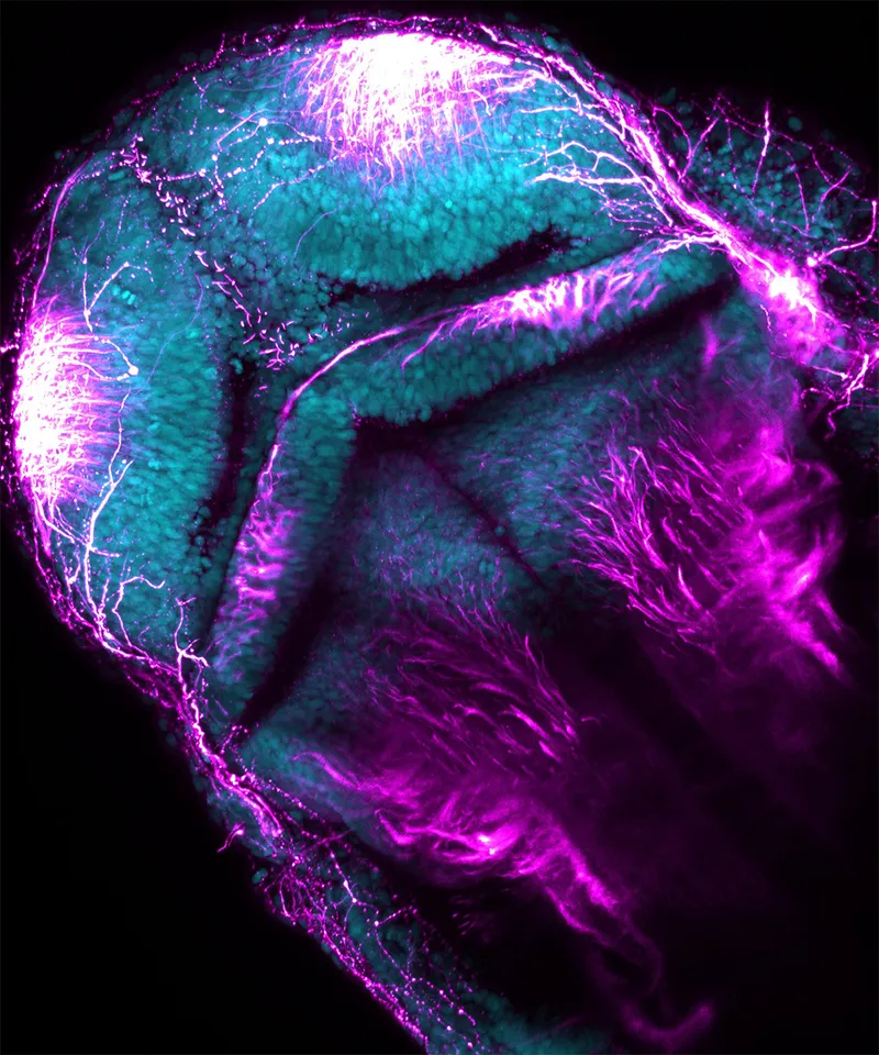
This image shows the rigid and unchanging tubular organisation of stabilised microtubules (blue) in a zebrafish central nervous system. Microtubules are tubular structures which form key components of the cytoskeleton in cells. They are essential for cell structure, intracellular transport, cell division and migration. Photo by Sara Anuar/GOSH
Shortlisted – 3D modelling for safer neurosurgical planning
This image shows a printhead moving left to right creating an accurate 3D printed model of a patient’s brain blood vessels. Arteriovenous malformations (AVM) are complex lesions that require extensive pre-surgical planning, and it is challenging for neurosurgeons to accurately visualise and deconstruct these structures. Photo by Luke Smith/GOSH
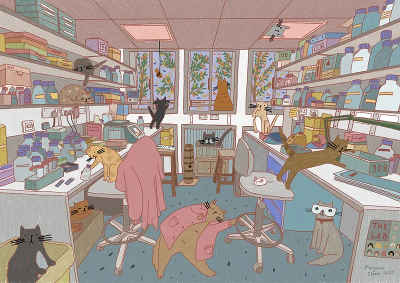
Shortlisted – Lab is home
The researcher Maryam used this illustration to show her tricky work-life balance, combining their second-home laboratory work with their home life – In this case her cats. Photo by Maryam Clark/GOSH
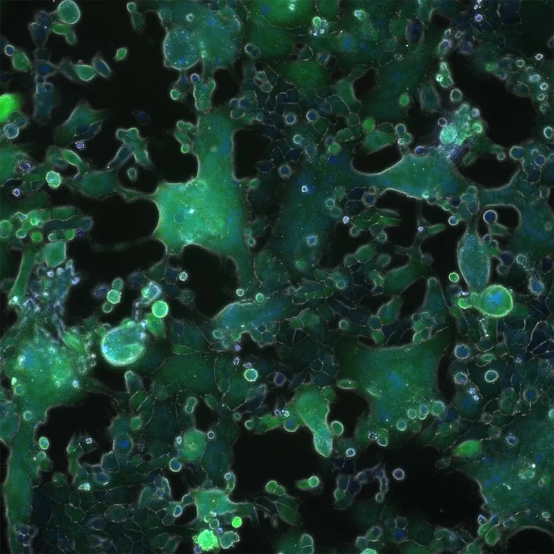
Shortlisted – Syncytial formation of RSV following cell infections
A cellular structure known as a syncytia is shown after being exposed to Respiratory Syncytial Virus (RSV). The green structures are RSV-infected cells and the blue circles show the cell nuclei. RSV is a respiratory virus that can cause mild flu-like symptoms and can, in some cases, lead to hospitalisation. Photo by Muhammad Pradhika Mapindra/GOSH
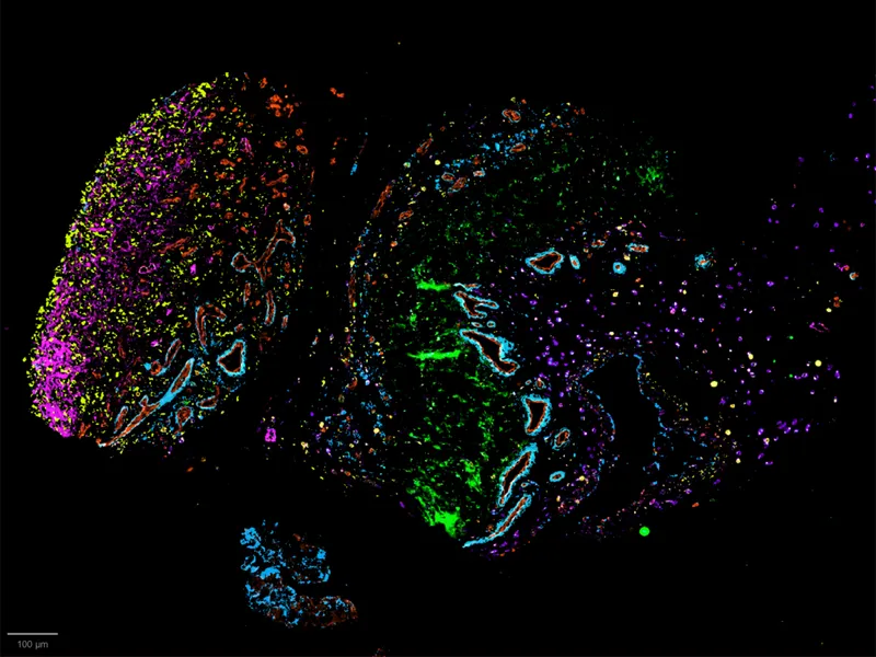
Shortlisted – Insights into Down's syndrome brain stem
Pink blood vessels and proliferative cells (blue/green) are imaged here in the hindbrain of a foetus with Down’s syndrome. The hindbrain, formed by the cerebellum and brainstem, regulates many vital functions including heart rate and breathing. Photo by Ekin Ucunco/GOSH
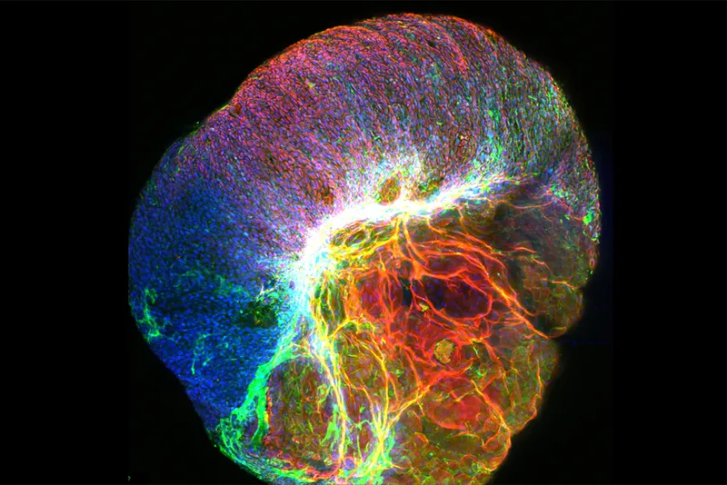
Panel favourite – What we 'kneed' to know
A knee joint of a four-year-old with arthritis. The red areas show the blood vessels infiltrating the tissue. They increase as the disease progresses, bringing in cells which attack the joint. The outer yellow and pink areas should be a thin barrier for the knee but becomes thicker as the joint tries to heal the damage caused by the inflammation. The green layers show scar tissue, which makes the joint stiffer and harder to move. Photo by Chrissy Bolton/Soren Lomholt/Patricia Reis-Nis/GOSH
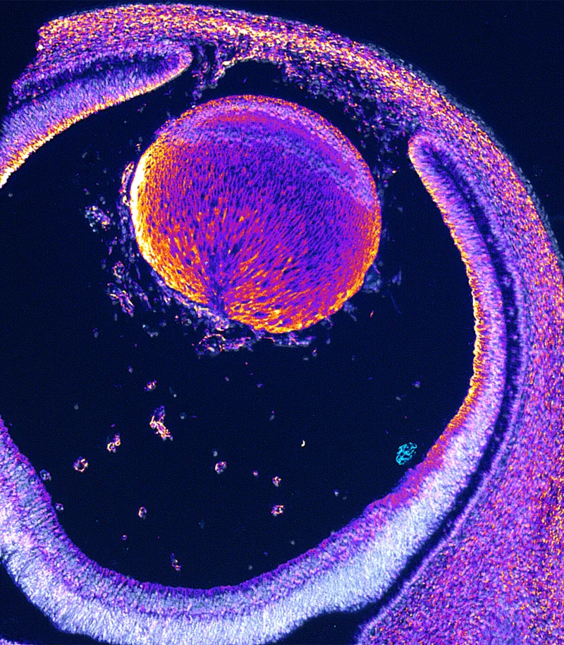
Panel favourite – It's all about seeing the world
An embryonic eye stained with dystrophin to visualise its location and its contribution to the developmental journey. This work aims to discover what dystrophin and its associated proteins look like in the developing brain and how it changes with development. Photo by Reem Alkharji/GOSH
Overall winner – The explosive potential of GI organoids
This image shows a gut ‘mini-organ’, known as an organoid. During a process used to visualise specific proteins, one of the organoids exploded, revealing its inner workings. Tiny organs like this one can be used to model gastrointestinal diseases, and are the perfect tool for scientists to study new therapies and test new drugs in the laboratory. Photo by Giada Benedetti/GOSH
James Cutmore is the picture editor of BBC Science Focus Magazine. He has worked on the magazine and website for over a decade, telling compelling science stories through the use of striking imagery. He holds a degree in Fine Art, and has been nominated for the British Society of Magazine Editors Talent Awards, being highly commended in 2020. His main areas of interest include photography that highlights positive technology and the natural world. For many years he was a judge for the Wellcome Trust's image competition, as well as judging for the Royal Photographic Society.
This website is owned and published by Our Media Ltd. www.ourmedia.co.uk