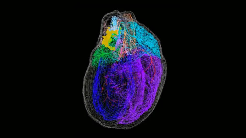
Most people tend to associate nerve cells (neurons) with the brain, but the heart also has a network of nerve cells known as the ‘intrinsic cardiac nervous system’ (ICN). Now, for the first time, a virtual 3D heart has been created that showcases the ICN on a cellular level.
It was created by scientists from diverse areas of study, and should allow researchers to precisely study the organisation and function of the heart’s neurons in incredible detail.
To create the 3D model, which was based on a rat heart, the scientists combined a variety of different techniques. First, they took thin slices through the length of the heart, obtaining microscope images and tissue samples in the process. They used these to create the foundation of the 3D construction.
Next, individual nerve cells were extracted from the rat hearts, and genetic information was obtained from them. This data was then fitted onto the 3D model, allowing the scientists to understand the role played by each nerve cell.

While the research was based on rat hearts, the team says that it should have an impact on human medicine.
For example, certain cases of heart disease have been improved by stimulating the vagus nerve, which runs all the way from the brain to the abdomen, branching off into the throat, heart, lungs and digestive system. Yet it is unclear why some heart disease cases can be cured in this way and others cannot.
Read more about the heart:
- Fitbit investigates whether wearables can diagnose heart conditions
- Revolutionary new valve could save children from repeated open-heart surgery
- Vegans and vegetarians at 'lower risk of heart disease', but have increased chance of stroke
“Evaluating these cardiac neurons from an anatomical and molecular perspective may help us better understand their function and develop therapies that can produce these protective effects of the vagus nerve onto the hearts of more patients,” said co-author Dr Jonathan Gorky, from Thomas Jefferson University.
The methods used to create this model have been made available via the SPARC programme at sparc.science, so that other teams can build upon the research, potentially leading the way for the development of virtual maps of other organs.
Reader Q&A: Why is the heart slightly to the left in the chest?
Asked by: Adam King, Huddersfield
The heart is located fairly centrally beneath the breastbone, but it does protrude towards the left. This is because the heart’s bottom-left chamber (the ‘left ventricle’) is responsible for pumping oxygenated blood around the whole body, so it needs to be stronger and larger than the right ventricle, which only pumps blood to the lungs. It’s this left ventricle that you can feel beating in your chest.
One in 10,000 people actually have a mirror-image heart which points towards the right – a condition known as ‘dextrocardia’.
Read more:
