
Have you ever wondered what a lab-grown organoid looks like, how your ears detect sound or what an aspirin tablet reveals under extreme magnification?
Well, we have the answers thanks to a selection of extraordinary science and health images submitted to the Wellcome Photography Prize.
In 2027, this prize will celebrate its 30th year of bringing science to life through photography, so we have gathered some of the most amazing images from the last 29 years.
All of them showcase the forefront of scientific discovery. Of the nine images here, two were taken by legendary photographer Spike Walker, the prize’s most prolific winner, who died in 2025. Spike had his work recognised 29 times over 12 years by Wellcome.
Catherine Draycott, who originally founded the prize, paid tribute to Walker at the launch of this year's competition.
She said: “Spike brought specimens alive with his skill, enthusiasm and creative techniques. From pond water to old microscope slides, he made incredibly vivid and arresting images that always excite curiosity and wonder.”
The submission window for next year’s prize is now closed, with the winners being announced at the 30th anniversary, in summer 2027.
Brain organoid
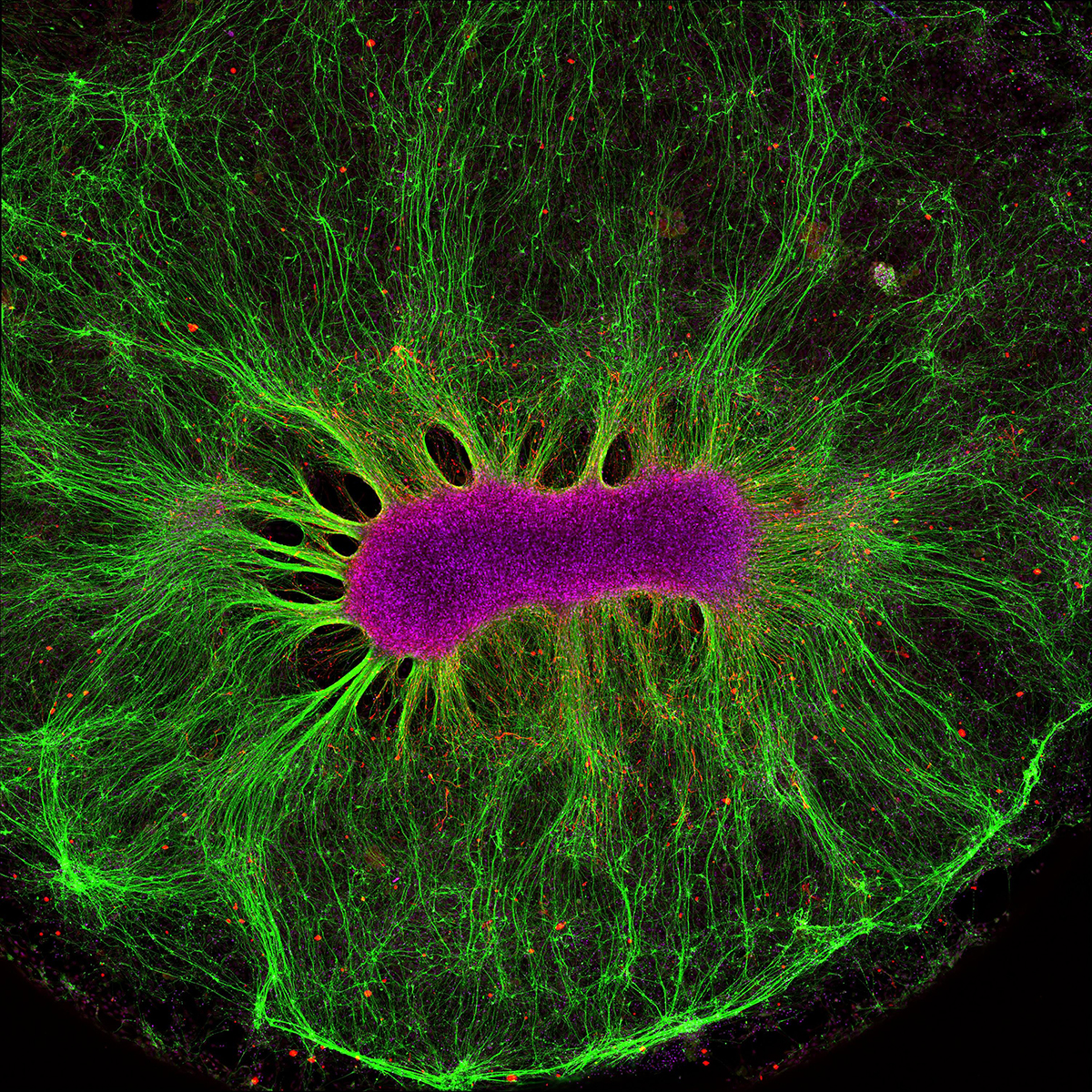
This image shows a cluster of brain cells grown in a lab on a synthetic polyethylene glycol (PEG) hydrogel, forming a structure known as an organoid.
From a central group of cells, nerve-like fibres grow outwards in all directions, forming a network. The image shows how brain cells can build complex patterns outside the body when given the right environment.
Paracetamol crystals
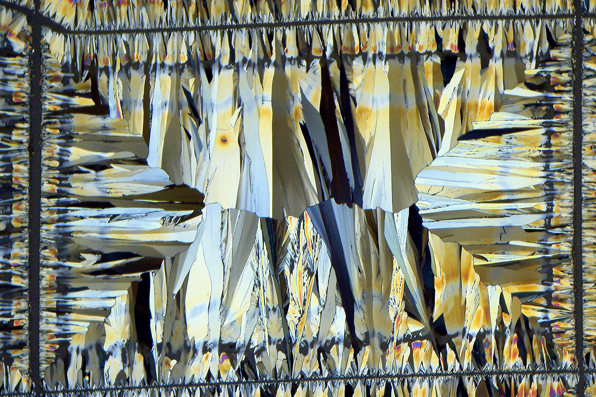
Paracetamol (acetaminophen) is one of the most widely used over-the-counter medicines for relieving mild to moderate pain and reducing fever. It is commonly taken for headaches, muscle aches, arthritis and cold or flu symptoms.
Despite more than a century of use, exactly how it works is only now being understood. Research led by the University of Copenhagen in 2025 has begun to uncover how acetaminophen acts on the central nervous system and interacts with receptors in the spinal cord.
The time of coronavirus

In this image, volunteers from the Blue Sky Rescue Team, the largest humanitarian NGO in China, are disinfecting the Qintai Grand Theatre in Wuhan, where COVID-19 was first identified.
Aspirin crystals
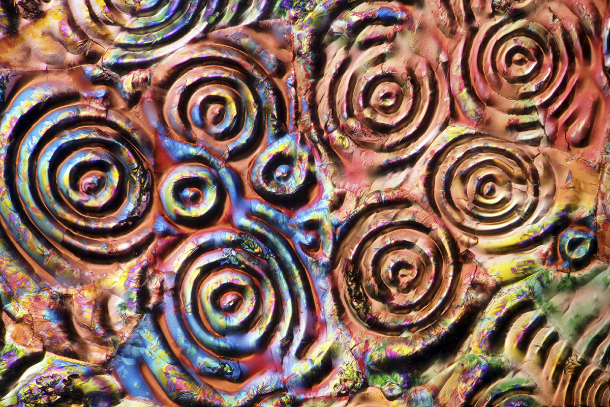
Aspirin crystals imaged using polarising light microscopy. These crystals can vary in size from nanometres to several millimetres.
Cochlea
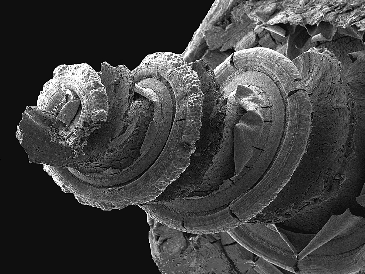
A scanning electron micrograph (SEM) of the inside of a guinea pig’s inner ear showing the hearing organ, or cochlea (left). Along the spiral structure is the organ of Corti (right) that contains rows of sensory hair cells that respond to different frequencies of sound. The cells send impulses along the auditory nerve to the brain. This image was entered into the inaugural 1997 competition.
Zebrafish embryos
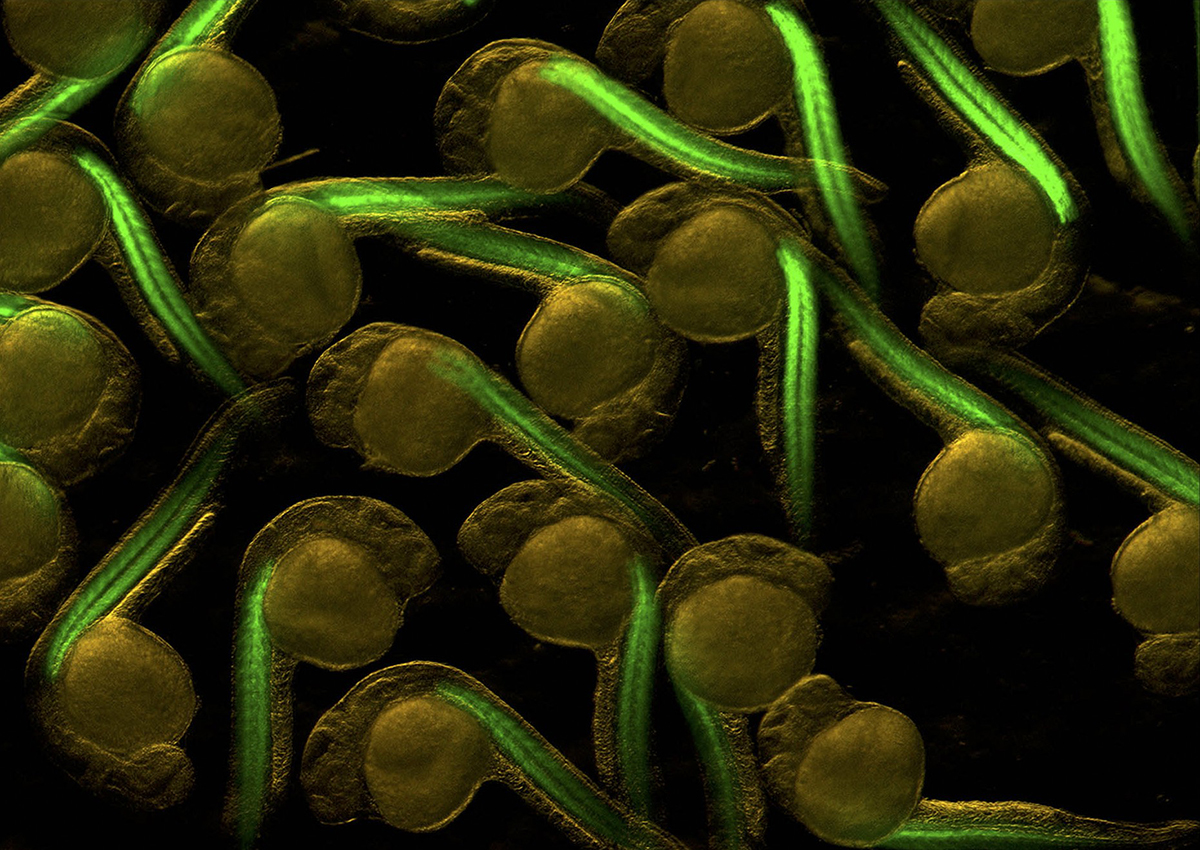
Zebrafish embryos with green fluorescent muscle blocks (myotomes). Imaged using fluorescence microscopy. Each one-day-old embryo in this image is approx 1mm (0.04 inches) wide.
Breast cancer cells treated with nano-sized drug carriers
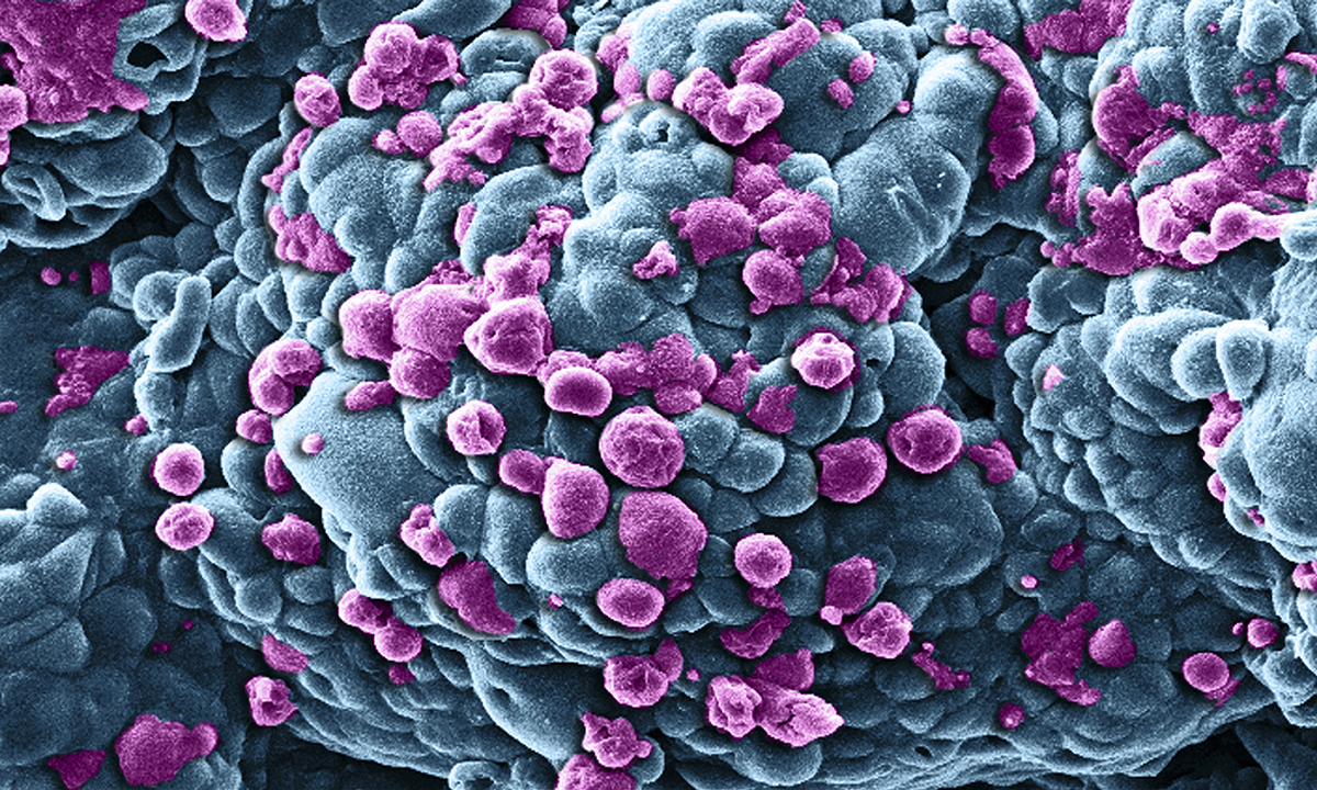
Breast cancer cells (blue) being treated with nano-sized drug carriers that are causing tumour cells to die (purple).
Mechanical heart
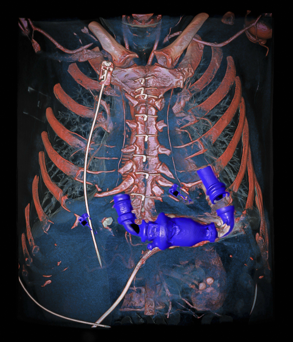
Dual-energy computed tomography angiography (DECT) of a human chest. The scan was performed with an extremely low radiation dose on a patient who had received a mechanical heart pump (blue) while waiting for a heart transplant.
The image was rendered with a 3D volume-rendering technique to check the wiring of the pump to the left side of the diseased heart and to the aorta.
Villi in the small intestine
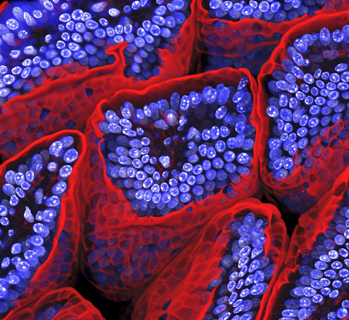
Villi, which facilitate digestion in mammals, are imaged here lining the small intestine of a mouse.
Read more:
