The winners and runners-up of this year's British Heart Foundation Reflections of Research image competition have just been announced, with a remarkable image of blood vessels in the lung being awarded the top prize. The winning photograph resembles an astronomical constellation, but instead of stars in the galaxy, tiny immune cells are scattered throughout blood vessels.
The winning photographer was Dr Régis Joulia, a BHF Research Fellow at the BHF Centre of Research Excellence at Imperial College London. His research looks at how the activation of immune cells disrupts the structure of lung blood vessels in chronic inflammatory conditions, such as pulmonary hypertension and asthma.
The British Heart Foundation is a charity that funds ground-breaking research that aims to get us closer to a world free from the fear of heart and circulatory diseases.
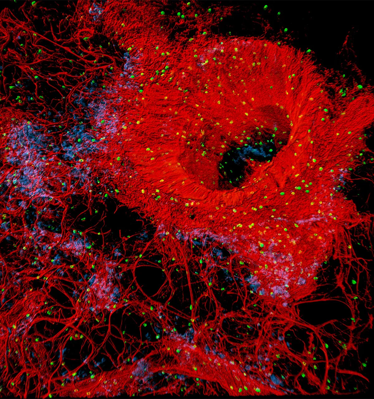
Overall winner - A flare of stellar vessels
This image shows a small region of a human lung and its rich supply of blood vessels in red. The tiny green dots are immune cells called mast cells in the lungs, and the blue dots are pericytes, which are crucial cells for blood vessels to maintain their structure.Photo by Dr Regis Joulia/British Heart Foundation Reflections of Research
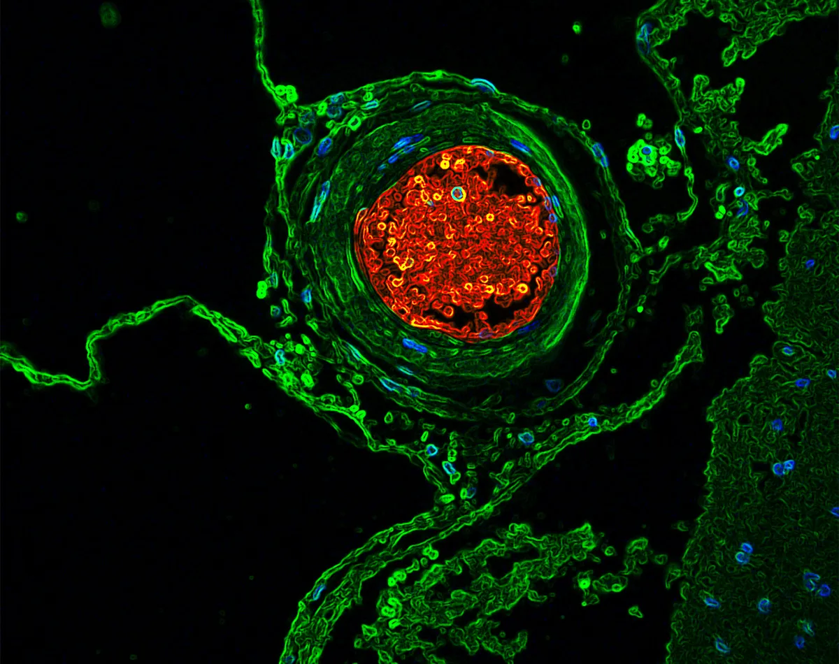
Joint runner-up - Blood vessel volcano
A human blood vessel is shown in green, with blood flowing through its centre in red. Arteries and veins are the highways of the body, responsible for transporting blood, oxygen and nutrients. By looking at the vessel architecture of people with and without hypertension, researchers hope to identify a new protein involved in high blood pressure. Photo by Dr Rhéure Alves-Lopes/British Heart Foundation Reflections of Research
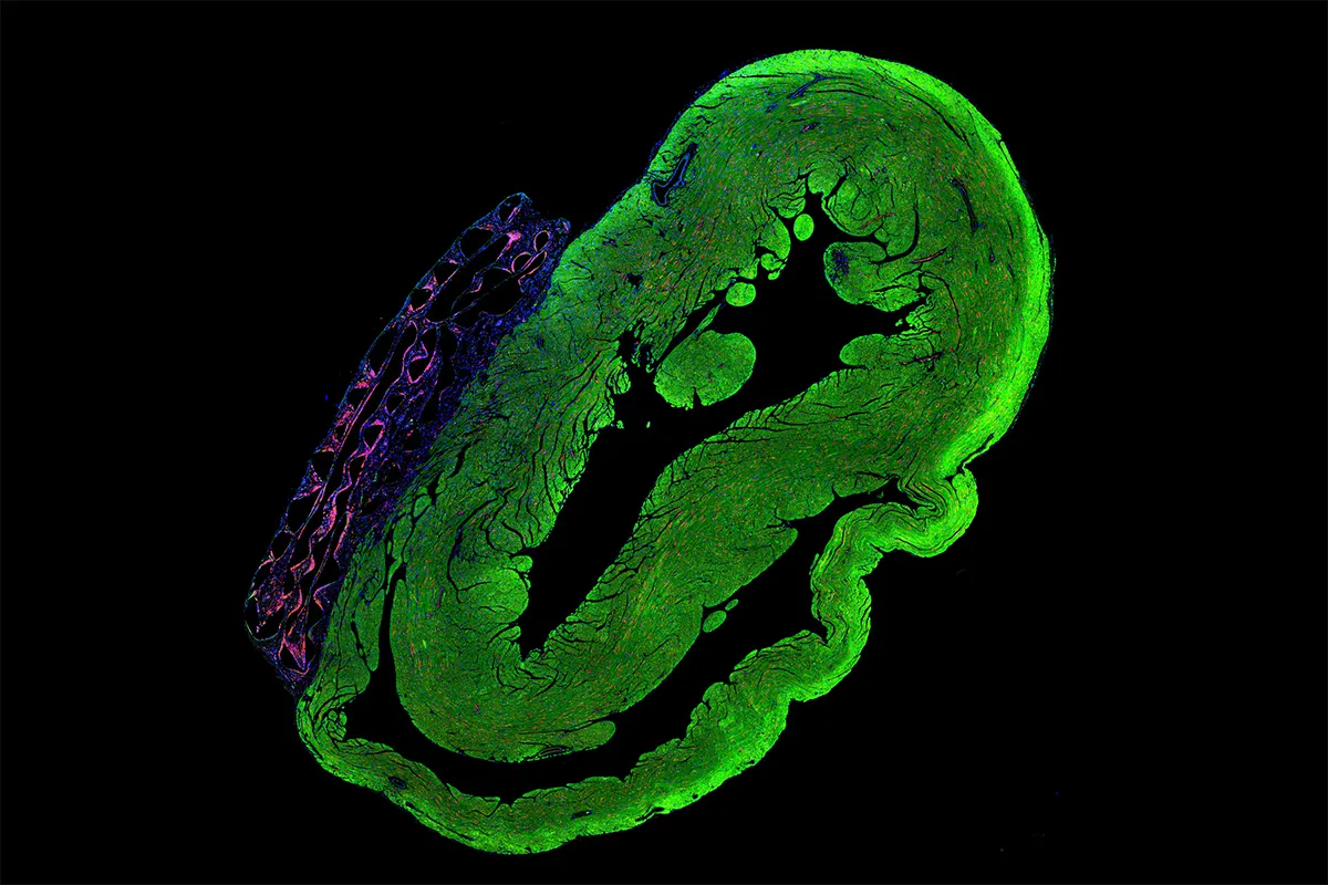
Joint runner-up - Regenerating heart
The green area of this image is a cross-section of a rat’s heart, with a patch of living heart cells shown in purple. This patch is made from stem cells, and acts as a sticking plaster to repair damage to the heart muscle after a heart attack. This repairable patch is being developed to replace and repair the cells lost after a heart attack, and to prevent heart failure from developing. Photo by Pragati Pandey/British Heart Foundation Reflections of Research
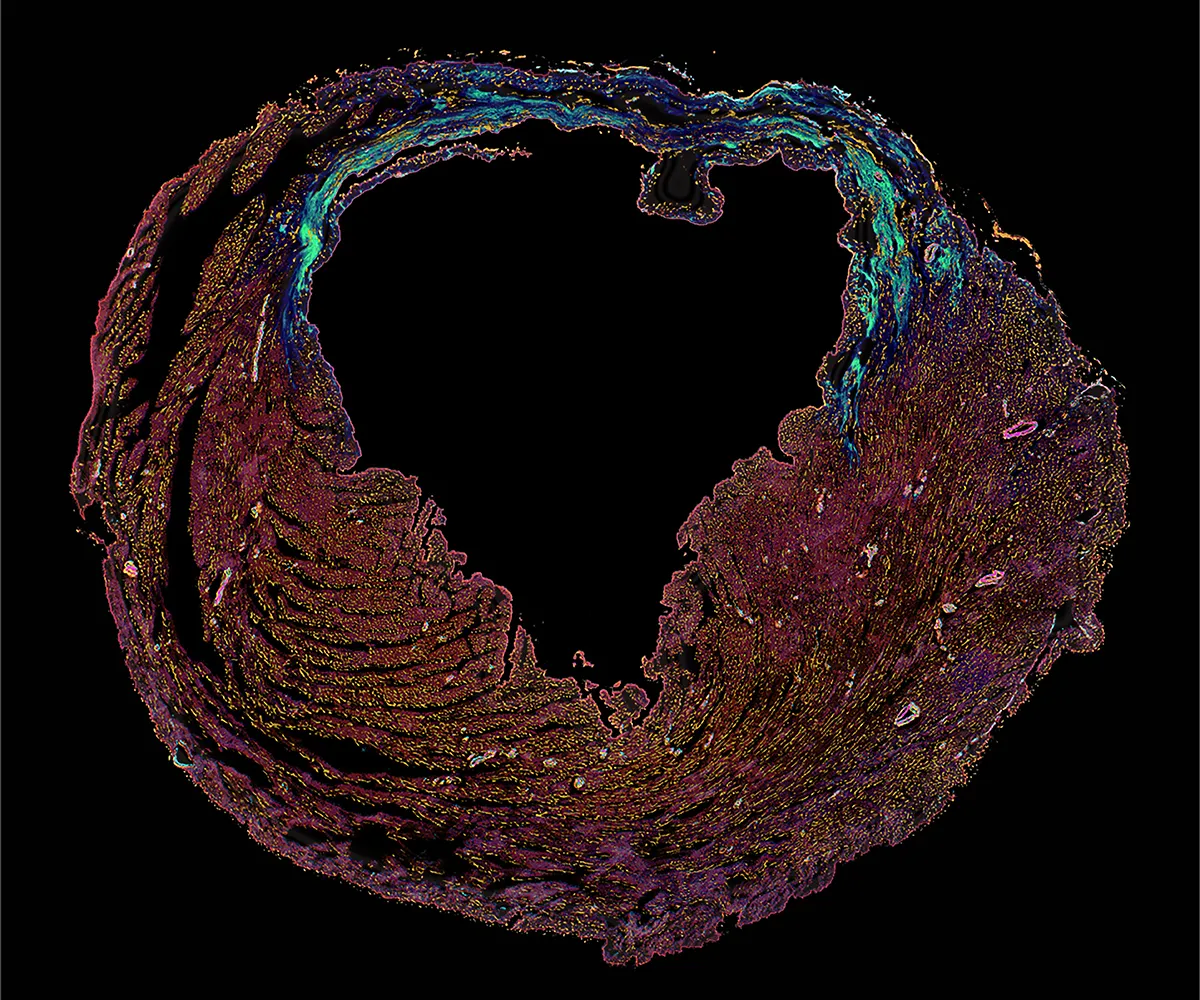
Joint runner-up - Heart within a heart
Damage to a heart following a heart attack. The death of heart muscle cells dramatically changed the structure of this heart’s main pumping chamber, which has caused it to become deformed into a cartoon heart shape. Photo by Christina Gkantsinikoudi/Dr Neil Dufton/British Heart Foundation Reflections of Research
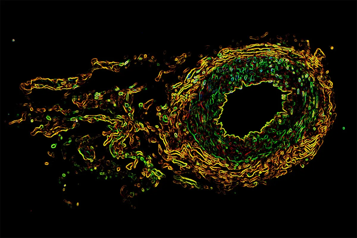
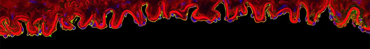
Shortlisted - Blood vessel comet
This is a human blood vessel taken from a biopsy. It shows the beautiful layers of different cells within the vessel. The black hole in the middle is where blood flows through, surrounded by a lining of endothelial cells captured in yellow. Next, glowing in green is the smooth muscle cells that are responsible for the vessel to widen and narrow. The outer yellow layer represents the adventitia, which contains connective tissue and nerves.Dr Karla Neves/British Heart Foundation Reflections of Research
Shortlisted - A heart in the lungs
This purple-grey heart shape in the middle of the image is an airway in the centre of a mouse lung, and the tiny air sacs called alveoli that form a mesh-like network around the airway are shown in pale purple. Surrounding the airway are large blood vessels highlighted in bright purple. Photo by Manpreet Kaur/British Heart Foundation Reflections of Research
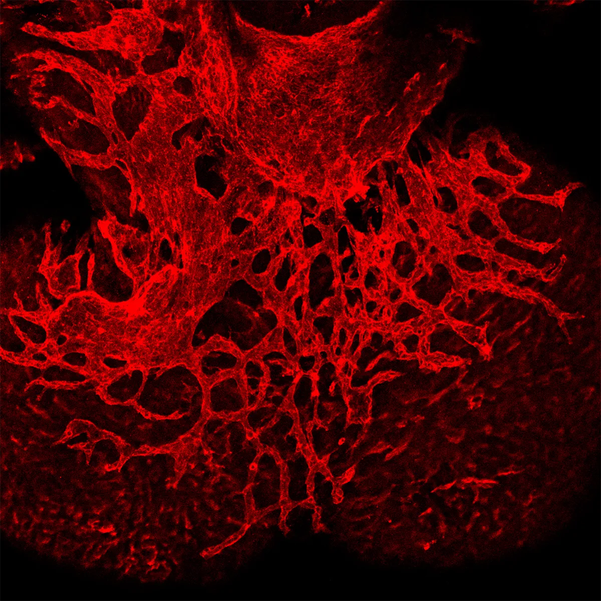
Shortlisted - Branching blood vessels in the heart
What looks like tree roots branching out into the soil at first glance, is in fact blood vessels growing on the outside surface of the heart - also known as the epicardium. Photo by Dr Joaquim Miguel Vieira/British Heart Foundation Reflections of Research
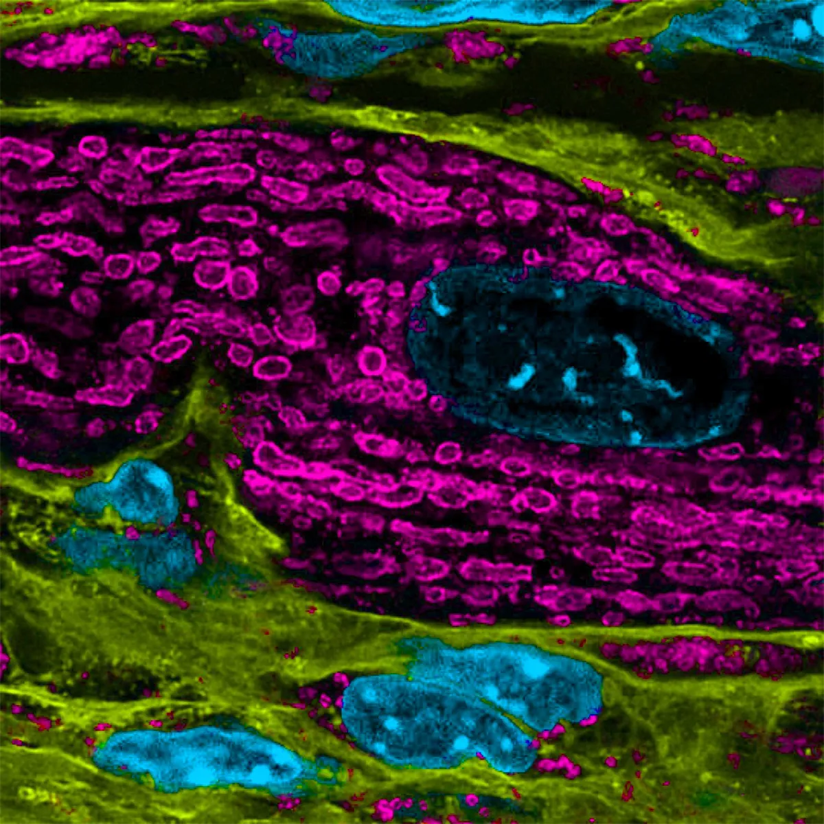
This colourful arrangement of shapes is the inner workings of a heart muscle cell, which has been affected by a heart condition called mitochondria-mediated cardiomyopathy. Mitochondria (shown in pink) are the engines of heart cells responsible for producing energy to power the beating motion of the heart. This image also reveals the nuclei of the heart cells in blue, which hold genetic information, and areas of heart scarring and immune cells in green.Photo by Jacky Fung/Amalia Sintou/British Heart Foundation Reflections of Research
Shortlisted - The inner lining of blood vessels
These undulating ripples of fluorescent ribbon are the lining of a blood vessel, with the black space showing where the blood flows inside the vessel. The bright red line is an elastic layer, which allows the blood vessels to contract and relax, and the blue dots reveal DNA. The bright green dye shows the glycocalyx - a gel-like layer that acts as a barrier between the blood flowing through the vessels and the cells. Photo by Nathalie Tarassova/British Heart Foundation Reflections of Research
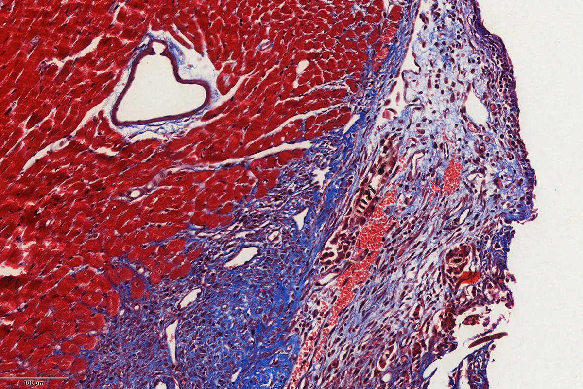
Shortlisted - A heart fighting fibrosis
This heart, floating in a sea of red and blue, is a small blood vessel supplying the heart muscle with oxygen. During a heart attack, these vessels become blocked. This starves part of the heart muscle of nutrients and oxygen which leads to damage and scarring in the heart muscle, a process called fibrosis. Excessive scarring reduces the heart’s ability to pump blood as well as it needs to. The image shows healthy heart tissue in red and scar tissue at the area of injury in blue.Photo by Georgios Kremastiotis/British Heart Foundation Reflections of Research
James Cutmore is the picture editor of BBC Science Focus Magazine. He has worked on the magazine and website for over a decade, telling compelling science stories through the use of striking imagery. He holds a degree in Fine Art, and has been nominated for the British Society of Magazine Editors Talent Awards, being highly commended in 2020. His main areas of interest include photography that highlights positive technology and the natural world. For many years he was a judge for the Wellcome Trust's image competition, as well as judging for the Royal Photographic Society.