This year's winners are Dr Daniyal Jafree, Radu Polschi, Aleksandra Letunovska and Reem Al-Saadi, for their image 'A 3D snapshot of the hidden highways in childhood kidney cancer'.
“I’ve been working at UCL Great Ormond Street Institute of Child Health for 7 years, and in that time, two things have become very clear." Dr Jafree tellsBBC Science Focus. "Firstly, it’s so important that we communicate our research to the people we ultimately hope to benefit: patients, their families and the public."
"Secondly, none of our research would be possible without a big team of scientists and clinical professionals, all with different backgrounds and experiences, all sharing the common goal of improving diagnosis and treatment for childhood diseases and cancer."
Other stand-out images include human nose cells infected with COVID, and an AI program that can detect wrist fractures in X-rays using the latest technology.
Great Ormond Street Hospital,with its team of dedicated paediatrichealthcare specialists, is one of the world’s leading children’s hospitals. Their pioneering research and cutting-edge treatments give hope to children with the rarest, most complex and often life-threatening conditions.
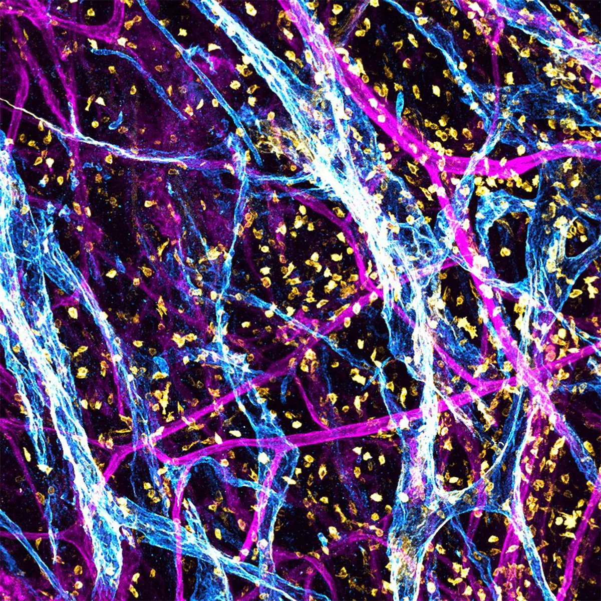
Overall winner - Hidden highways in childhood kidney cancer
Blue lymph networks amongst blood vessels (in purple) and immune cells (in yellow). The lymph network is responsible for waste disposal in healthy cells, but they can be ‘hijacked’ by cancer cells and then be used to spread the tumour cells around the body. Photo by Dr Daniyal Jafree/Radu Polschi/Aleksandra Letunovska/Reem Al-Saadi/Great Ormond Street Hospital
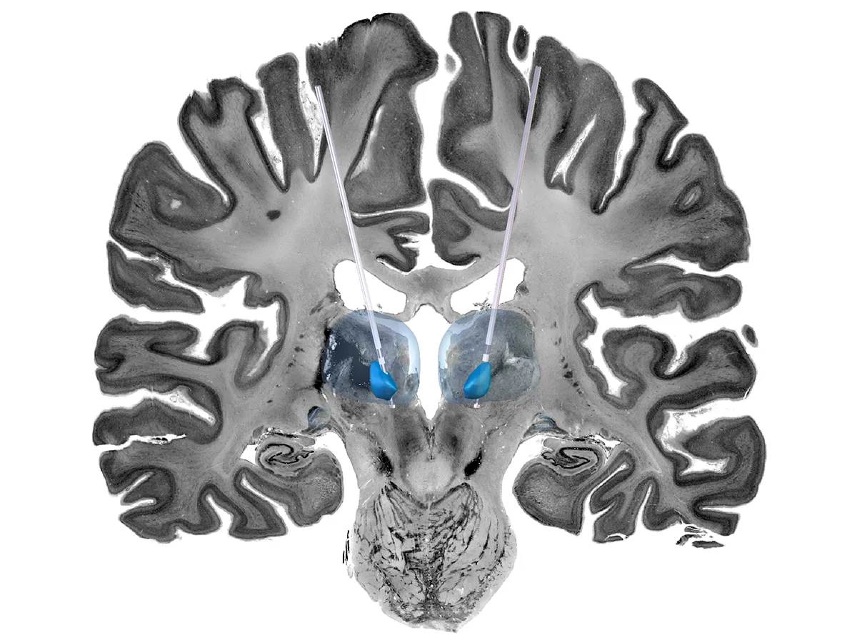
Top 3 - Stimulating the brain
This image has captured the cross-section of a simulated brain, used to help study potential treatments for epilepsy in children.Deep brain stimulation (DBS) is a type of brain surgery that uses electrical pulses to change the way the brain works. Here, a representative cross-section of the brain (looking from front to back) shows two DBS wires implanted in the thalamic regions (shown in blue). These regions are the parts of the brain involved in the routing of electrical signals.Photo by Rory Piper/Lewis Spitz/Great Ormond Street Hospital
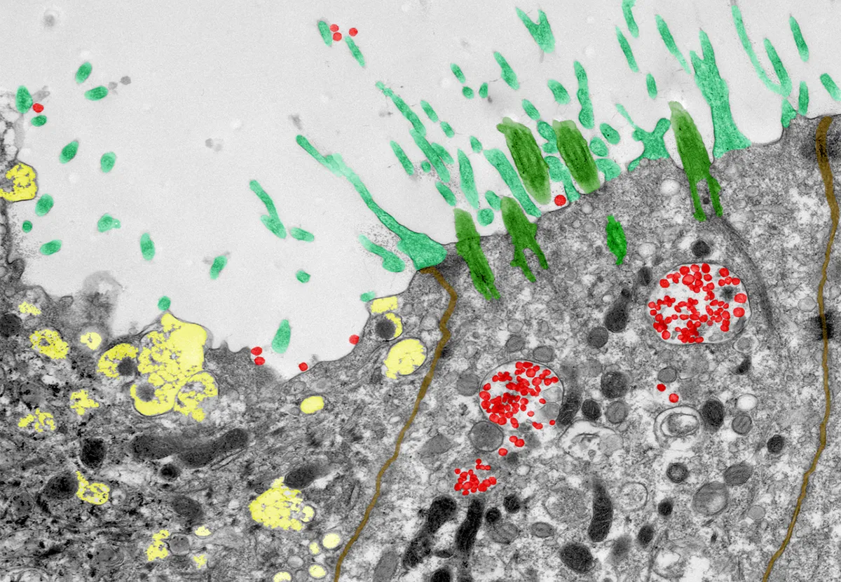
Top 3 - Nasal COVID-19 factories
This striking collection of shapes and colours shows human nose cells infected with SARS-CoV-2 (the viral particles are shown in red). It was taken as part of a study investigating how COVID infection differs with age. Photo by Dr Maximillian Woodall/Dr Samuel Ellis/Dr Andreia Pinto/Great Ormond Street Hospital
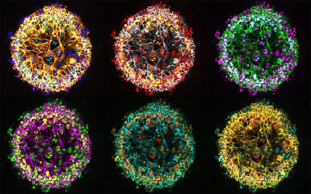
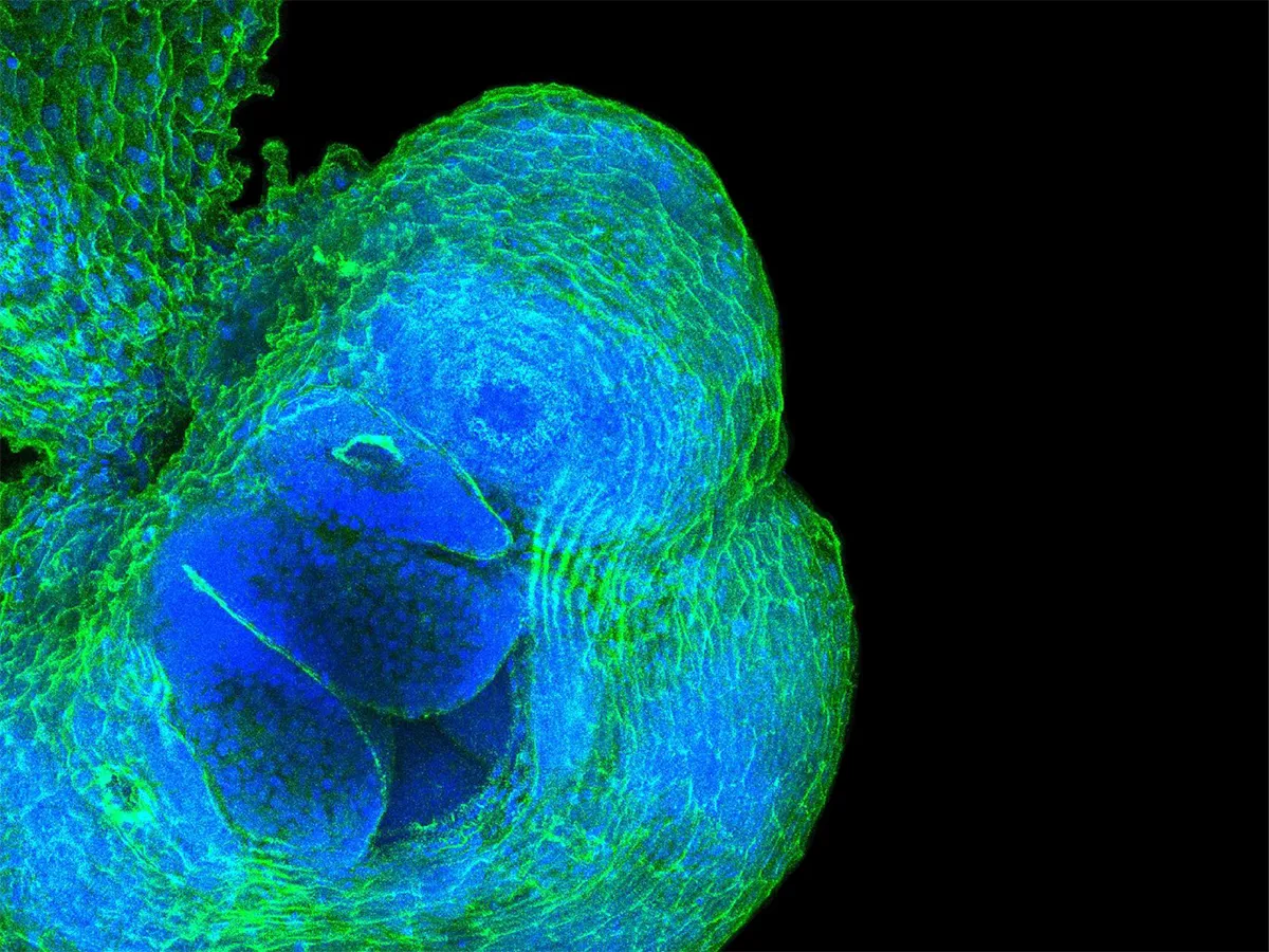
Shortlisted - Cellular system
These twinkling spheres are in fact developing embryos on glass beads.In order to understand changes in the embryo that may lead to developmental conditions in babies (spina bifida, for example), researchers must mimic the changing shape of organs. This image shows how cells that develop into the brain and spinal cord (neuroepithelial cells) grow on beads and mimic the curved shape of the head region.Photo by Makis Ampartzidis/Great Ormond Street Hospital
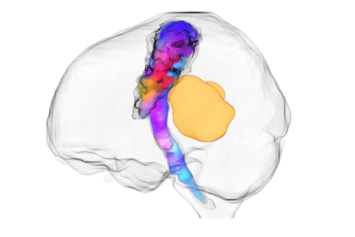
Shortlisted - Neurone bundle and brain tumour
This image is a visualisation of the interaction between a brain tumour (orange) and a bundle of nerve fibres produced using magnetic resonance imaging (MRI). The watercolour-stained ‘bundle’ shows the pathway that carries signals from the brain to the spinal cord, to control movement on the right side of the body.This image was created using a new technique, called diffusion MRI, that will hopefully make it easier and faster for neurosurgical teams to use MRI to improve surgical outcomes, even without highly specialist MRI skills.Photo by Fiona Young/Great Ormond Street Hospital
Shortlisted - Polarity proteins
This luminescent pool of blue and greens shows how a specific protein is involved in the maintenance of the shape and structure of cells in the body. This is known as cell polarity. This protein is used to study the development of the brain in zebrafish.Photo by Sara Anuar/Great Ormond Street Hospital
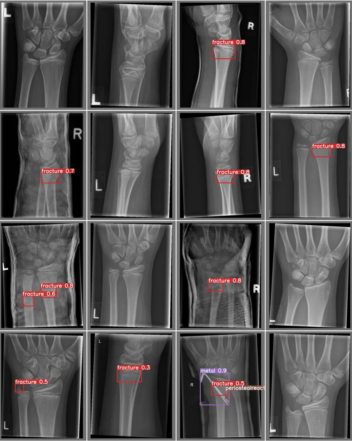
Shortlisted - Artificial Intelligence Detection of Wrist Fractures
This image is a collage of X-rays of children who have potentially broken bones in their wrist. The boxes in some of the images highlight abnormalities in these X-rays picked up by artificial intelligence (AI) programmes.Trained AI programmes have the potential to spot fractures like a radiologist, but in milliseconds. Combining the expertise of trained healthcare professionals and powerful AI programmes could reduce the number of missed fractures and ensure children get the right treatment quickly.Photo by Cato Pauling/Great Ormond Street Hospital
Shortlisted - Modern art in kidney biology
This image shows the lymphatic system of an adult mouse kidney, which is very similar to a human kidney. The lymphatics are a draining network that help the body remove unwanted fluids and molecules from tissue. The system is also vital for the movement of immune cells throughout our body.This image was taken as part of research to understand how a certain protein is turned on and off across this system within the kidney and how that may impact inflammation in kidney disease.Photo by Eva-Maria Funk/Great Ormond Street Hospital
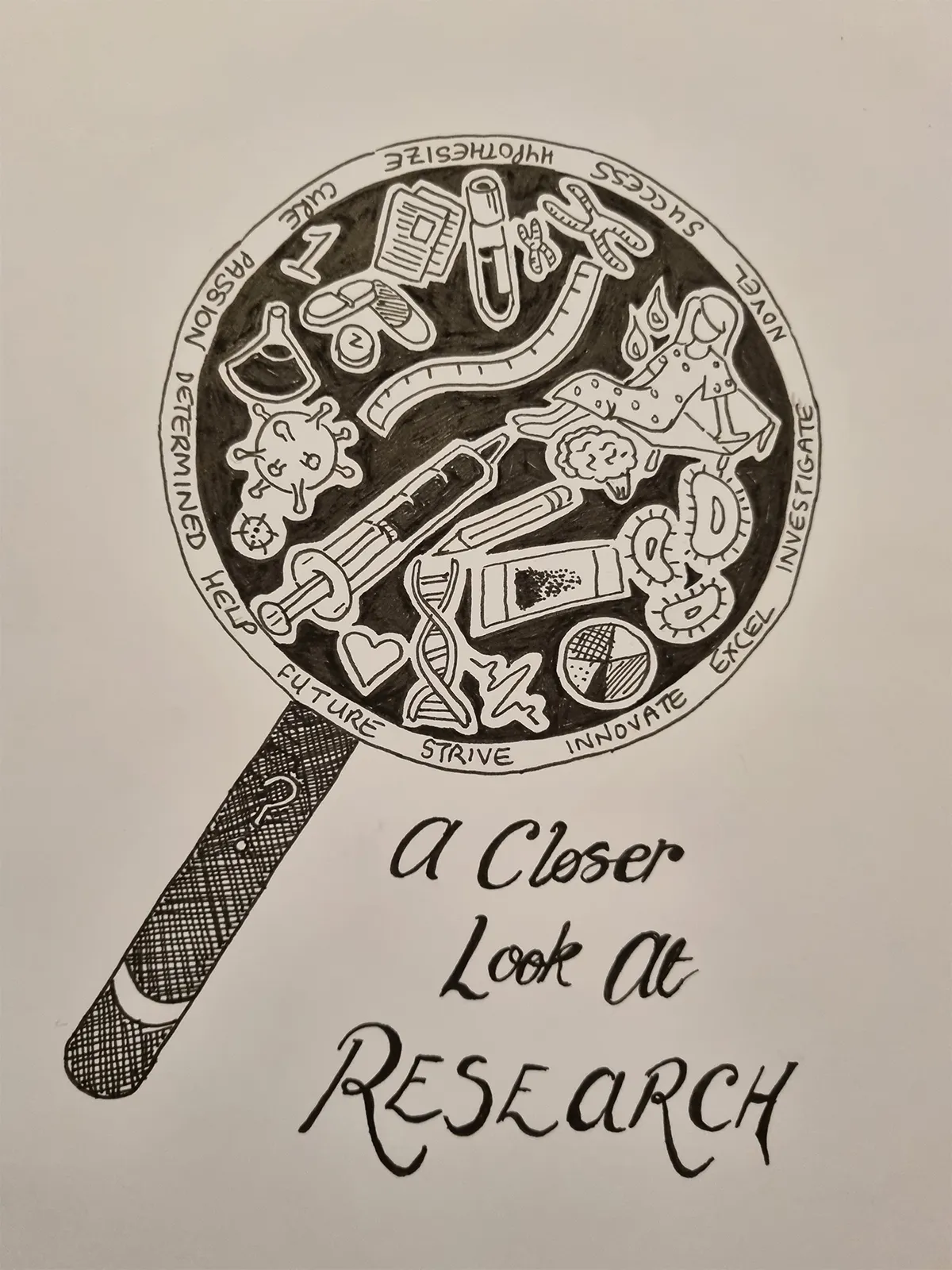
Shortlisted - A closer look at research
This illustration aims to communicate what is involved in development and innovation in healthcare. Research is only possible through the passion, hard work and expertise of everyone involved.Photo by Jade Sugars/Great Ormond Street Hospital
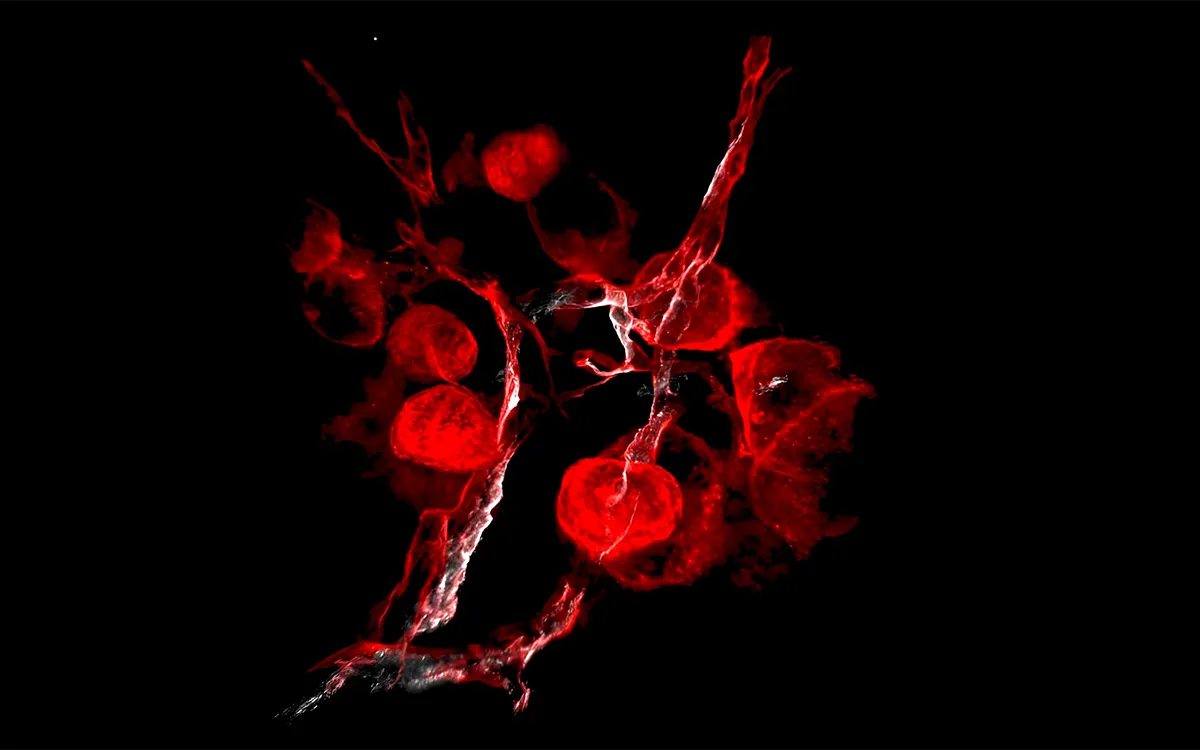
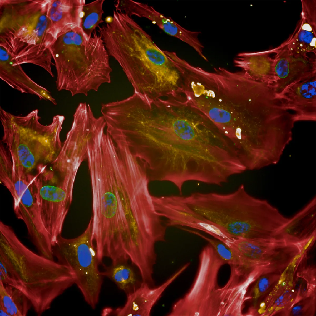
Shortlisted - Astrocytes
These red cells are known as astrocytes, named because of their star-like shape. These cells play an important role in the brain alongside nerve cells.These astrocytes were grown from stem cells that were taken from a young patient with genetic epilepsy. Scientists can use cells like these to understand how astrocytes from patients with epilepsy differ from those without the condition.Photo by Dr Jenny Lange/Great Ormond Street Hospital
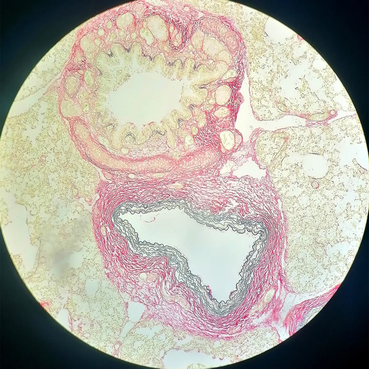
Shortlisted - Gaseous exchange
Resembling the cratered surface of a moon, this image shows a microscope image of a lung biopsy.The top structure shows an air sac, and below it a blood vessel. Air sacs and blood vessels work in combination to allow oxygen to enter the blood, and carbon dioxide to leave it. The biopsy was stained with a chemical which highlights blood vessels and the elastin protein in found in the tissue. These stains are vital in diagnostic tests to help teams identify changes in the lung tissue.Photo by Lucien Bonfante/Great Ormond Street Hospital
James Cutmore is the picture editor of BBC Science Focus Magazine. He has worked on the magazine and website for over a decade, telling compelling science stories through the use of striking imagery. He holds a degree in Fine Art, and has been nominated for the British Society of Magazine Editors Talent Awards, being highly commended in 2020. His main areas of interest include photography that highlights positive technology and the natural world. For many years he was a judge for the Wellcome Trust's image competition, as well as judging for the Royal Photographic Society.